. 2014 Nov-Dec;21(6):555-66.
doi: 10.1111/xen.12125. Epub 2014 Sep 1.

Development of a consensus protocol to quantify primate anti-non-Gal xenoreactive antibodies using pig aortic endothelial cells

Affiliations

Development of a consensus protocol to quantify primate anti-non-Gal xenoreactive antibodies using pig aortic endothelial cells

Agnes M Azimzadeh et al. Xenotransplantation. 2014 Nov-Dec.

Abstract

Background: Scientists working in the field of xenotransplantation do not employ a uniform method to measure and report natural and induced antibody responses to non-Galα(1,3)Gal (non-Gal) epitopes. Such humoral responses are thought to be particularly pathogenic after transplantation of vascularized GalTKO pig organs and having a more uniform assay and reporting format would greatly facilitate comparisons between laboratories.

Methods: Flow cytometry allows examination of antibody reactivity to intact antigens in their natural location and conformation on cell membranes. We have established a simple and reproducible flow cytometric assay to detect antibodies specific for non-Gal pig antigens using primary porcine aortic endothelial cells (pAECs) and cell culture-adapted pAEC cell lines generated from wild type and α1,3galactosyl transferase knockout (GalTKO) swine.

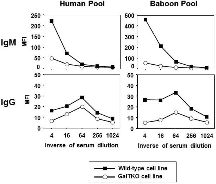
Results: The consensus protocol we propose here is based on procedures routinely used in four xenotransplantation centers and was independently evaluated at three sites using shared cells and serum samples. Our observation support use of the cell culture-adapted GalTKO pAEC KO:15502 cells as a routine method to determine the reactivity of anti-non-Gal antibodies in human and baboon serum.

Conclusions: We have developed an assay that allows the detection of natural and induced non-Gal xenoreactive antibodies present in human or baboon serum in a reliable and consistent manner. This consensus assay and format for reporting the data should be accessible to laboratories and will be useful for assessing experimental results between multiple research centers. Adopting this assay and format for reporting the data should facilitate the detection, monitoring, and detailed characterization of non-Gal antibody responses.

Keywords: antibody; cell line; galactosyl transferase; non-Gal antibody; xenoreactive assay; xenotransplantation.


Figures

Figure 1
Figure 1. Phenotypic characterization of WT and GalTKO porcine endothelial cell lines (KO:15502 and WT:14259)
A. Expression of αGal was assessed using FITC-conjugated lectin BS-I-B4. B. Staining for CD31. Unstained cells were used as negative control for IB4 lectin staining, and an isotype-matched control was used for CD31 staining (grey).
Figure 2
Figure 2. IgM and IgG antibody binding to WT and GalTKO pAEC cell lines
Serial dilutions (1:4 to 1:1024) of naïve human and baboon serum pools were tested using the standardized protocol described in the Methods section and Table 1. Data were expressed as median fluorescence intensity (MFI).
Figure 3
Figure 3. Inter-laboratory evaluation of the consensus protocol
The figure compared IgM (A) and IgG (B) reactivity for naïve human and baboon serum pools and serum from baboons sensitized by cardiac xenotransplantation (BS1 and BS2) tested using the pAEC line KO:15502 cells as measured in the three test sites. In panel B, median fluorescence intensity (MFI) units (top panel) were converted to relative fluorescence units (RFU) as a ratio of MFI to the secondary only control (RFU no serum, middle panel) or to the human pool (RFU hu pool, bottom panel), using the mathematical equations listed at the bottom. BS1, BS2: serum samples collected from baboons sensitized to pig antigens by a GalKO heart xenograft in absence of immunosuppression as indicated in Table 4.
Figure 4
Figure 4. Sources of variability
A, Comparison of results produced using different flow cytometers (FACs Calibur or FACs Verse), using MFI or RFU data analysis. B, Compares the effects of cell culture conditions on MFI values for IgM (left) or IgG (right) antibody reactivity. BS3: Serum sample collected from a baboon sensitized to pig antigens by a GalTKO heart xenograft in absence of immunosuppression as indicated in Table 4.
Figure 5
Figure 5. Comparison of antibody reactivity to primary and cell culture adapted GalTKO cell lines
A, The reactivity of naïve human serum was tested using the pAEC line KO:15502 (red) or primary pAECs from 3 individual GalTKO pigs (black lines); B, Serum samples from baboons sensitized by cardiac xenotransplantation (BS4 - 6) were tested using the pAEC line KO:15502 (red), primary pAECs from the donor pig (blue), or primary pAECs from unrelated third-party pigs (black). Since results were expressed as MFI or RFU, legend for the Y axis was omitted for clarity and units of measure are indicated on the top of each figure panel. The results from three representative animals are shown. BS4 - 6: Serum samples collected from baboons sensitized to pig antigens by a GalTKO heart xenograft with immunosuppression as indicated in Table 4.

References

    1. Ekser B, Ezzelarab M, Hara H, et al. Clinical xenotransplantation: the next medical revolution? Lancet. 2012;379:672–683. - PubMed
    1. Pierson RN, III, Dorling A, Ayares D, et al. Current status of xenotransplantation and prospects for clinical application. Xenotransplantation. 2009;16:263–280. - PMC - PubMed
    1. Allan JS. Understanding xenotransplantation risks from nonhuman primate retroviruses. Curr Top Microbiol Immunol. 2003;278:101–23. 101–123. - PubMed
    1. Issa NC, Wilkinson RA, Griesemer A, et al. Absence of replication of porcine endogenous retrovirus and porcine lymphotropic herpesvirus type 1 with prolonged pig cell microchimerism after pig-to-baboon xenotransplantation. J Virol. 2008;82:12441–12448. - PMC - PubMed
    1. Scobie L, Padler-Karavani V, Le Bas-Bernardet S, et al. Long-term IgG response to porcine Neu5Gc antigens without transmission of PERV in burn patients treated with porcine skin xenografts. J Immun. 2013;191:2907–2915. - PMC - PubMed

Publication types