Review
. 2014 Nov-Dec;3(6):403-18.
doi: 10.1002/wdev.147. Epub 2014 Aug 29.

Development and evolution of the pharyngeal apparatus

Affiliations
Review

Development and evolution of the pharyngeal apparatus

Aude Frisdal et al. Wiley Interdiscip Rev Dev Biol. 2014 Nov-Dec.

Abstract

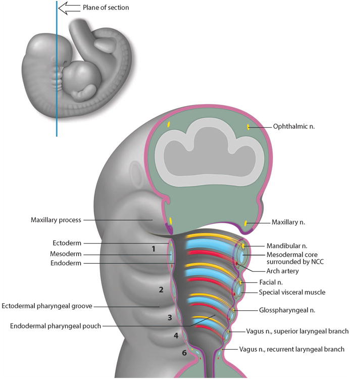
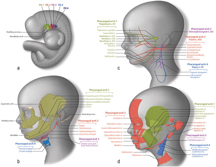
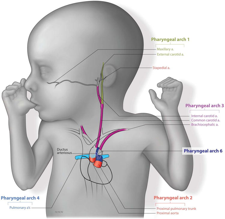
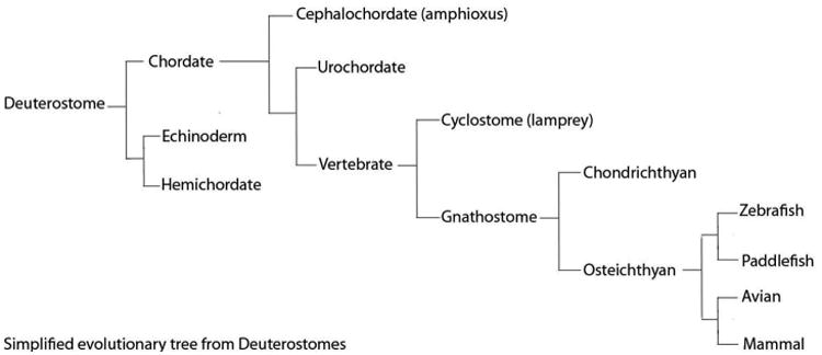
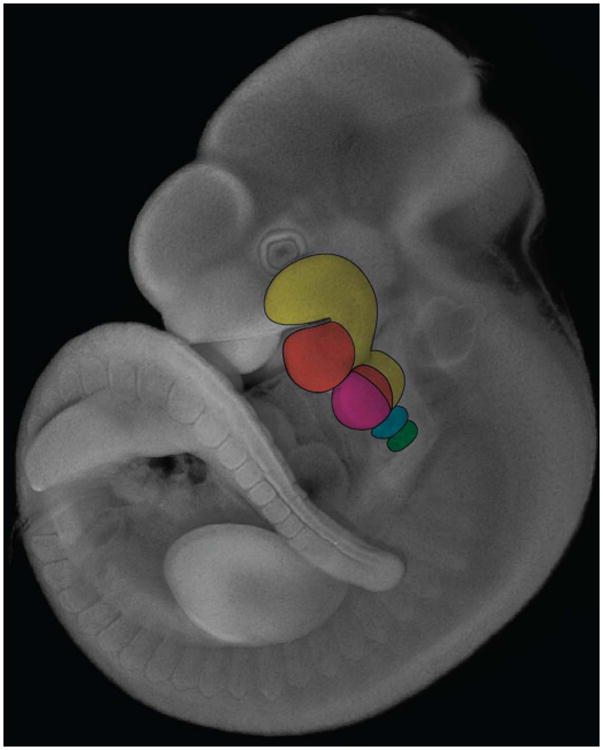
The oral or pharyngeal apparatus facilitates the dual functions of respiration and feeding. It develops during embryogenesis from transient structures called pharyngeal arches (PAs), which comprise a reiterated series of outgrowths on the lateral side of the head. The PAs and their segmental arrangement are highly conserved throughout evolution from invertebrate chordates such as amphioxus, through to vertebrate agnathans including avians, squamates, and mammals. The structural organization of the PAs is also highly conserved and involves contributions from each of the three primary endoderm, mesoderm, and ectoderm germ layers. The endoderm is particularly important for PA formation and segmentation and also plays a critical role in tissue-specific differentiation. The ectoderm gives rise to neural crest cells (NCC) which provide an additional layer of complexity to PA development and differentiation in vertebrates compared to invertebrate chordates that do not possess NCC. Collectively, the PAs give rise to much of the neurovasculature and musculoskeletal systems in the head and neck. The complexity of development renders the pharyngeal apparatus prone to perturbation and subsequently the pathogenesis of birth defects. Hence it is important to understand the signals and mechanisms that govern the development and evolution of the pharyngeal complex.


Figures

Figure 1
Figure 1. Structural Organization of the Pharyngeal Arches in a Human Embryo
Schematic illustration of the internal organization of the developing PAs. Each PA is numbered from 1 to 6. PAs are lined externally by ectoderm in pink and internally by endoderm in purple. Inside, each PA is a mesenchymal core (blue) composed of mesoderm cells surrounded by NCC cells alongside the nerve (yellow) and blood vessel (red) components.
Figure 2
Figure 2. Pharyngeal Arch Differentiation
Figure 2a: Schematic illustration of the pharyngeal region of a human embryo. Figure 2b: Skeletal derivatives of the NCC. Bones of the face are formed by two ossification processes. Endochondral bones are formed via a cartilage template and include the maleus (from Meckel's cartilage), and the incus (from maxillary cartilage) from the 1st PA (green). The stapes, the styloid process and lesser horn of hyoid (from Reichter's cartilage) come from the 2nd PA (red). Dermal bone formation consists of direct ossification of the NCC without a cartilage intermediate. Dermal elements include the squamous part of the temporal bone, the zygomatic, the maxillary bone and the mandible, which are derived from 1st PA (green). Figure 2c: Neural derivatives of the NCC The trigeminal nerve (V) comprises 3 subdivisions: the ophthalmic nerve, the maxillary nerve and the mandibular nerve and develops in association with the 1st PA, (green). The facial nerve invades the 2nd PA, (red). The glossopharyngeal invades the 3rd PA, (purple) and the vagus nerve the 4th PA, (dark blue). Figure 2d: Muscular derivatives of the mesoderm. Muscles (green) of mastication are derived from mesoderm of the 1st PA. Muscles of facial expression (red) are derived from 2nd PA mesoderm. The stylopharyngeus (purple) is the only muscle that is derived from the 3rd PA mesoderm. The external muscles of the pharynx (blue) are derived from 4th PA mesoderm.
Figure 3
Figure 3. Establishment of the musculature, vasculature, and PNS of the face in mouse embryo
a. LacZ staining marking NCC. b. tbx1 in situ hybridization showing the myogenic core of each PA. c. Pecam immunostaining marking endothelial cells of the developing vasculature. d.Tuj1 immunostaining marking NCC and placode-derived neurons, e. Eya2 in situ hybridization, delimiting the cranial placodes.
Figure 4
Figure 4. Pharyngeal Arch Vascular Patterning
The maxillary artery (green) is derived from the 1st PAA. The stapedial (red) is derived from the 2nd PAA. The carotid system (purple) derives from the 3rd PAA. The left 4th PAA gives rise to the aortic arch while the right PAA gives rise to the subclavian artery and contributes to the pulmonary arteries (light blue). Finally, the 6th PAA give rise to the ductus arteriosus (dark blue) and the pulmonary arteries (light blue).
Figure 5
Figure 5. Evolutionary Cladogram
Evolutionary tree showing the classification of species from deuterostomes to mammals. NCCs appear during the transition to vertebrates.
Figure 6
Figure 6. Dlx and Hox gene control of pharyngeal patterning
Dapi staining of E10.5 mouse embryo. Expression domain of Dlx1 and Dlx2 (yellow) in the 1st and 2nd PA. Nested expression of Dlx5 and Dlx6 (orange) within the Dlx1 and Dlx2 domains of expression. Hox2 group genes are expressed in the 2nd PA (pink) and more posterior Pas, while Hox group 3 (blue) and Hox group 4 (green) genes are expressed in the 3rd and 4th PAs resepectively.

References

    1. Jones NC, Trainor PA. The therapeutic potential of stem cells in the treatment of craniofacial abnormalities. Expert Opin Biol Ther. 2004;4:645–657. - PubMed
    1. Scambler PJ. The 22q11 deletion syndromes. Hum Mol Genet. 2000;9:2421–2426. - PubMed
    1. Walker MB, Trainor PA. Craniofacial malformations: intrinsic vs extrinsic neural crest cell defects in Treacher Collins and 22q11 deletion syndromes. Clin Genet. 2006;69:471–479. - PubMed
    1. Sperber GH. Craniofacial development. 2001
    1. Le Douarin NMK. The Neural Crest. 1999

Publication types

LinkOut - more resources