

Retinal and choroidal thickness measurements using spectral domain optical coherence tomography in anterior and intermediate uveitis
- PMID: 25176513
- PMCID: PMC4236668
- DOI: 10.1186/1471-2415-14-103
Retinal and choroidal thickness measurements using spectral domain optical coherence tomography in anterior and intermediate uveitis
Abstract
Background: Macular edema is a common cause of visual loss at uveitic patients. The aim of our study was to investigate retinal and choroidal thickness at the macula in anterior (AU) and intermediate (IMU) uveitis and in healthy individuals using spectral domain optical coherence tomography (SD-OCT).
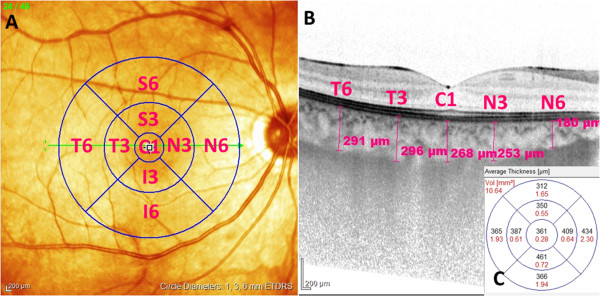
Methods: Case-control study of 21 patients with AU and 23 patients with IMU and 34 age-matched healthy controls was performed with Spectralis SD-OCT (Heidelberg Engineering, Germany). High resolution SD-OCT scans and macular mapping were applied for automated measurement of retinal thickness. Standardized, masked manual measurement of the choroidal thickness was performed in the center of the ETDRS fields on enhanced depth imaging (EDI) scans. Evaluation of central retinal subfield thickness, 3 mm and 6 mm perifoveal rings was performed in the corresponding ETDRS zones in patient groups.
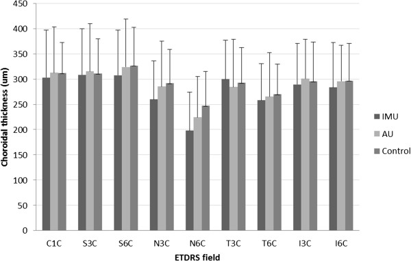
Results: The mean central retinal subfield thickness was significantly higher in IMU (368.65 ± 115.88 μm, p = 0.0003), but not significantly different in AU (290.42 ± 26.37 μm p = 0.6617) compared to that of in controls (278.55 ± 18.31 μm). In both uveitis groups retina was significantly thicker in the 3 and 6 mm perifoveal rings than that of in controls (359 ± 15.24 μm in AU and 390.55 ± 70.90 μm in IMU vs 345,41 ± 15.28 μm in the control group, p = 0.0388 and p < 0.0001) in the 3 mm and (313.83 ± 16.63 μm in AU and 343.33 ± 57.29 μm in IMU vs 299 ± 13.82 μm in the control group, p = 0.0171 and p < 0.0001) in the 6 mm ring. Central choroidal thickness was 311.94 ± 60.48 μm in the control eyes, showed no significant difference in AU (312.61 ± 90.35 μm) and IMU (303.17 ± 93.66 μm) eyes, and was also similar at the perifoveal rings.
Conclusion: Significant topographical changes could be detected in the macula of AU and IMU patients. Retinal thickness in the perifoveal rings was increased both in AU and IMU, but in the center only in IMU. Choroidal thickness seems to be unaffected by uveitis, even in the presence of macular edema, at least in the early stage of the inflammatory disease process.
Figures

References
-
- Shulman S, Goldenberg D, Habot-Wilner Z, Goldstein M, Neudorfer M. Optical coherence tomography characteristics of eyes with acute anterior uveitis. Isr Med Assoc J. 2012;14:543–546. - PubMed
Publication types
MeSH terms
LinkOut - more resources
Full Text Sources
Other Literature Sources
