. 2014 Sep 11;8(5):1484-96.
doi: 10.1016/j.celrep.2014.07.056. Epub 2014 Aug 28.

Dominant lethal pathologies in male mice engineered to contain an X-linked DUX4 transgene

Affiliations

Dominant lethal pathologies in male mice engineered to contain an X-linked DUX4 transgene

Abhijit Dandapat et al. Cell Rep. .

Abstract

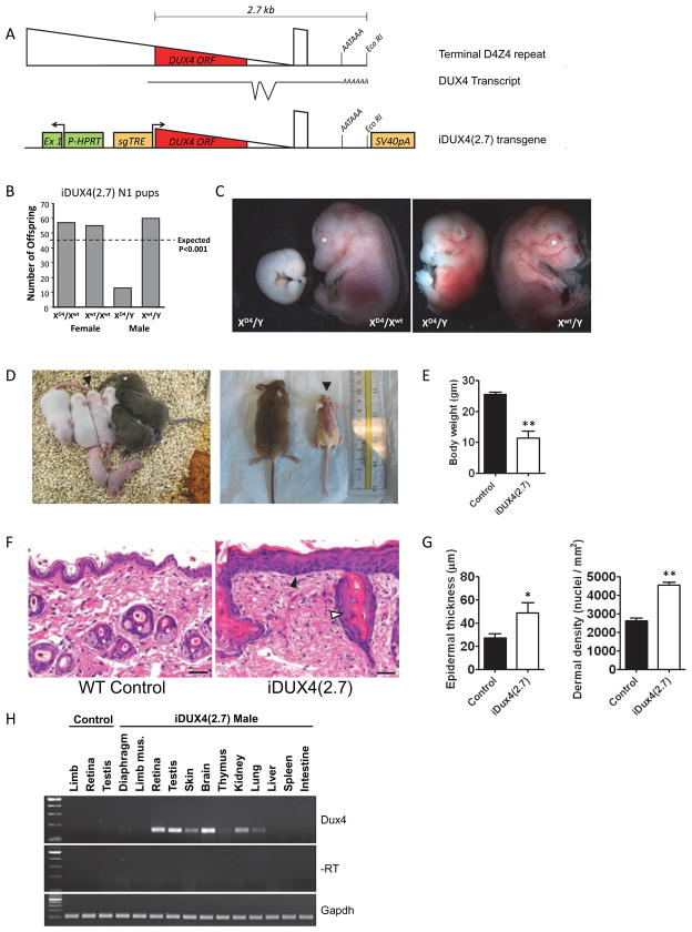
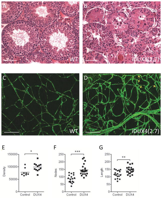
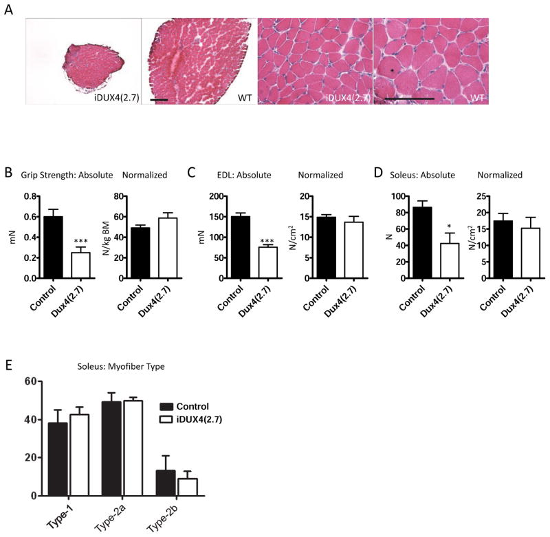
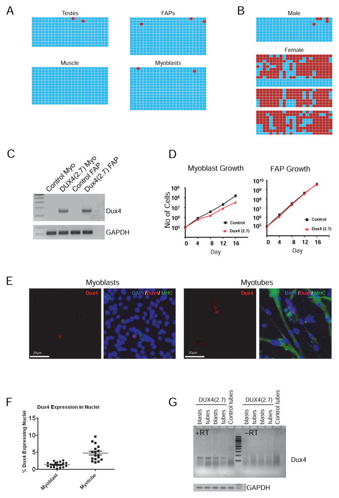
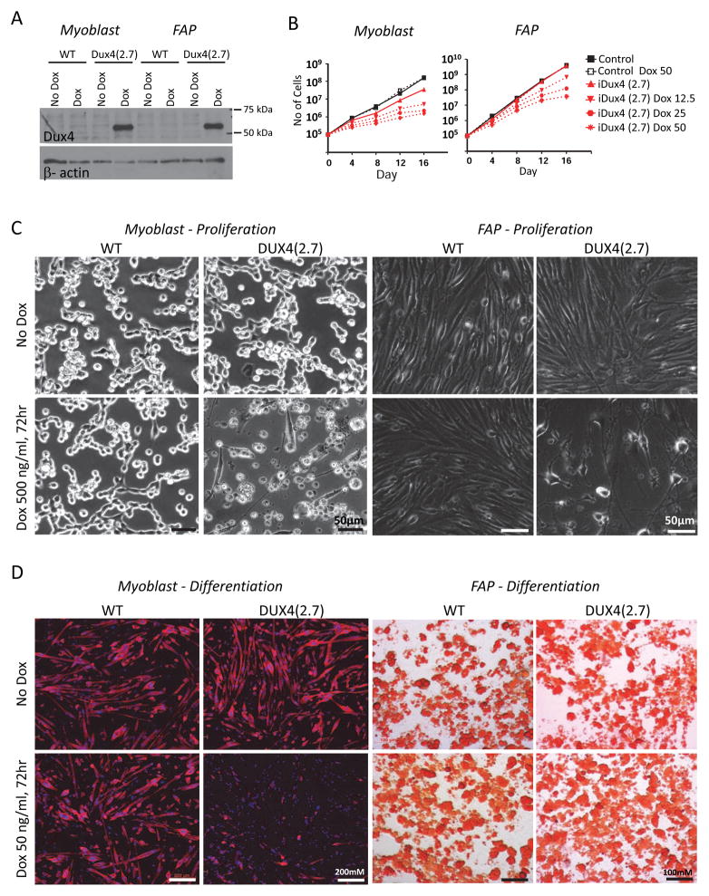
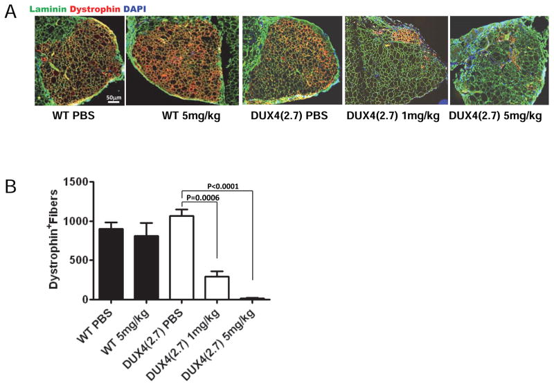
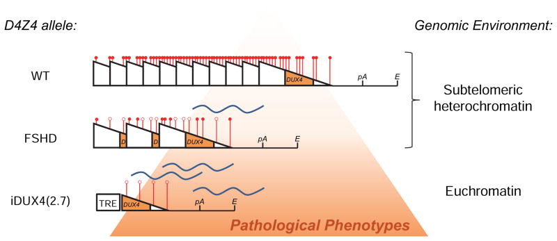
Facioscapulohumeral muscular dystrophy (FSHD) is an enigmatic disease associated with epigenetic alterations in the subtelomeric heterochromatin of the D4Z4 macrosatellite repeat. Each repeat unit encodes DUX4, a gene that is normally silent in most tissues. Besides muscular loss, most patients suffer retinal vascular telangiectasias. To generate an animal model, we introduced a doxycycline-inducible transgene encoding DUX4 and 3' genomic DNA into a euchromatic region of the mouse X chromosome. Without induction, DUX4 RNA was expressed at low levels in many tissues and animals displayed a variety of unexpected dominant leaky phenotypes, including male-specific lethality. Remarkably, rare live-born males expressed DUX4 RNA in the retina and presented a retinal vascular telangiectasia. By using doxycycline to induce DUX4 expression in satellite cells, we observed impaired myogenesis in vitro and in vivo. This mouse model, which shows pathologies due to FSHD-related D4Z4 sequences, is likely to be useful for testing anti-DUX4 therapies in FSHD.


Figures

Figure 1
Figure 1. The iDUX4(2.7) transgene is male-specific dominant lethal
(A) Schematics of the terminal D4Z4 repeat (above), the DUX4 transcript with introns and proposed AATAAA polyadenylation signal (below) and the construct integrated into the X chromosome (bottom). HPRT, hypoxanthine phosphor-ribosyl transferase; Ex1, exon 1, sgTRE, second generation tet-response element. (B) Genotypes of live pups from iDUX4(2.7) female carriers bred to WT males and chi-square P-values. (C) iDUX(2.7) embryos at E14.5. XD4 indicates the transgenic iDUX4(2.7)-bearing chromosome. Most male carrier embryos display severe growth delay or resorption. (D) Examples of litters with runted iDUX4(2.7) males. Left: a P5 litter with two obvious runts, and one animal, identified by arrowhead, with some hair loss that later became runted. Right: littermate WT and iDUX4(2.7) males at P28. (E) Weight at 6 weeks, p<0.05. (F) H&E staining of skin. Note the hypertrophic epithelium (filled arrowhead) and the abnormal glands (open arrowhead). Scale bar = 20 μm. (G) The epidermal thickness and nuclear density in the dermis were measured at 6 locations per section and averaged (* p<0.04; ** p<0.001) and the experiment was repeated with three mice. (H) RT-PCR-detection of the DUX4 transcript in tissues of iDUX4(2.7) transgenic male mice. Robust and repeatable expression were always seen in the brain, retina and testis. Other tissues displayed sporadic and weaker expression.
Figure 2
Figure 2. Phenotypes in testis and retina
(A) Cross section through WT seminiferous tubules. Scale bar = 50 μm. (B) Cross section through iDUX4(2.7) seminiferous tubules. (C) Vasculature in WT retina: montage of z-stack laser scanning confocal microscopy images of retina flat mounts stained with anti-CD31/PECAM showing, normal presentation of vasculature. Scale bar = 50 μm. (D) Vasculature in iDUX4(2.7) retina showing dense network of disorganized and twisted/looping vessels. Yellow arrows indicate twisted vessels. (E) Morphometric analysis of retinal images following skeletonization showing vascular density measured as immunoreactive pixels (p<0.05). (F) Morphometric quantification of vessel branching measured by nodes (p<0.001). (G) Morphometric quantification of total vessel length. For E–G, each point represents values/field, averaged from 3–4 different fields per retina; n=7 iDUX4, n=8 WT (p< 0.01).
Figure 3
Figure 3. Muscle in iDUX4(2.7) animals
(A) H+E staining of cross sections through soleus muscles of iDUX4(2.7) males (left) and male littermate controls (right). Scale bar is 100 μm (low magnification images) or 200 μm (high magnification images). (B) Grip strength measurements at 6 weeks, absolute at left (p<0.001), normalized to body mass at right (N=4). (C) Maximal isometric force generated by the extensor digitorum longus muscle at 6 weeks, absolute at left (p<0.001), normalized to cross sectional area at right (N=4). (D) Maximal isometric force generated by the soleus muscle at 6 weeks, absolute at left (p=0.026), normalized to cross sectional area at right (N=4). (E) Fiber-type analysis based on myosin heavy chain isoform expression.
Figure 4
Figure 4. Methylation and DUX4 expression in primary cells
(A) Methylation of the iDUX4(2.7) transgene in various tissues, and primary muscle cells. Bisulfite-treated DNA was amplified by PCR, cloned and sequenced. Blue boxes indicate unmethylated cytosines of CpG dinucleotides and red boxes indicate methylated cytocines along the PCR product. (B) Methylation of the transgene in male and female peripheral blood. One male aged 6 weeks, and three independent females aged 6, 9, and 36 weeks are shown. Methylated sequences in females were greatly overrepresented compared to the 1:1 ratio predicted by X-inactivation for a heterozygous female (p=0.0). (C) RT-PCR detection of the DUX4 transcript in proliferating myoblasts and FAPs. (D) Growth rate of iDUX4(2.7) myoblasts and FAPs compared to cells from WT littermate controls. Myoblasts displayed a significant growth disadvantage. No difference was seen in FAP growth rate. One experiment of 3 similar replicates is shown. (E) Immunofluorescent detection of DUX4 protein in myoblasts and myotubes. Sections are also stained with DAPI (blue) and antibody to myosin heavy chain (MHC, in green). (F) Quantification of DUX4+ nuclei, expressed as average percentage and SD of total per microscopic field, over 19 separate fields. One of 3 similar replicates is shown. (G) Comparison of levels of DUX4 expression by RT-PCR in myoblasts vs. myotubes. See also Figure S1.
Figure 5
Figure 5. Effects of induced DUX4 expression
(A) Western blot for DUX4 expression in proliferating myoblasts and FAPs exposed to a high dose (500 ng/mL) of dox. One representative blot of 3 independent experiments is shown. (B) Dox dose-response growth curves for myoblasts (left) and FAPs (right) exposed to very low doses of dox to induce DUX4 expression.Myoblasts displayed a more severe growth inhibition and greater sensitivity to dox. (C) Cellular morphology of proliferating myoblast (left) and FAP (right) cultures exposed to a high dose (500 ng/mL) of dox. (D) Myoblasts and FAPs exposed to a low dose (50 ng/mL) of dox and cultured under myogenic or adipogenic differentiation conditions, respectively. Myotubes (left) were stained for myosin heavy chain; adipocytes (right) were stained with Oil Red O. Note that low dose induction of DUX4 inhibits myogenic but not adipogenic differentiation.
Figure 6
Figure 6. DUX4 expression impairs myogenic regeneration in vivo
(A) Representative examples of TA muscles one month after transplantation of 1,800 iDUX4(2.7) satellite cells in the presence or absence of daily doxycycline injection (either 1 mg/kg or 5 mg/kg) to induce DUX4 or control carrier (PBS) injection. The overall muscle architecture is indicated by laminin staining (green). Donor cell contribution to new fibers is evaluated by counting dystrophin+ (red) fibers. Doxycycline treatment impaired contribution to myofiber regeneration in a dose-dependent manner. (B) Quantification of myofiber engraftment. For each group, N=6. Bars indicate SEM.
Figure 7
Figure 7. The iDUX4(2.7) allele vs. human D4Z4 alleles
D4Z4 is indicated by a triangle. Methylation state is indicated at 6 representative sites for each array, with a red ball indicating methylation and an open circle indicating lack of methylation. In humans, when D4Z4 is present in a large tandem array within subtelomeric heterochromatin at 4q, a repeat-induced silencing mechanism leads to heterochromatinization of the array (indicated by compaction of the triangles and their respective methylation marks) and hypermethylation of DNA (indicated by red circles), which effectively silences the locus. When the array number is reduced in an FSHD allele, a loss of repeat-induced silencing leads to an opening up of chromatin (more space between the triangles), and a relative demethylation, leading to some transcription of DUX4. In the iDUX4(2.7) mouse, the single copy (which is not subject to repeat-induced silencing) and location within euchromatin, results in even greater opening, a complete absence of methylation, and greater transcription / transcription in additional tissues, leading to a more severe, lethal, phenotype.

References

    1. Agha-Mohammadi S, O’Malley M, Etemad A, Wang Z, Xiao X, Lotze MT. Second-generation tetracycline-regulatable promoter: repositioned tet operator elements optimize transactivator synergy while shorter minimal promoter offers tight basal leakiness. J Gene Med. 2004;6:817–828. - PubMed
    1. Arpke RW, Darabi R, Mader TL, Zhang Y, Toyama A, Lonetree CL, Nash N, Lowe DA, Perlingeiro RC, Kyba M. A New Immuno- Dystrophin-Deficient Model, the NSG-Mdx Mouse, Provides Evidence for Functional Improvement Following Allogeneic Satellite Cell Transplantation. Stem Cells 2013 - PMC - PubMed
    1. Bakker E, Wijmenga C, Vossen RH, Padberg GW, Hewitt J, van der Wielen M, Rasmussen K, Frants RR. The FSHD-linked locus D4F104S1 (p13E-11) on 4q35 has a homologue on 10qter. Muscle Nerve. 1995;2:S39–44. - PubMed
    1. Block GJ, Narayanan D, Amell AM, Petek LM, Davidson KC, Bird TD, Tawil R, Moon RT, Miller DG. Wnt/beta-catenin signaling suppresses DUX4 expression and prevents apoptosis of FSHD muscle cells. Hum Mol Genet. 2013;22:4661–4672. - PMC - PubMed
    1. Bosnakovski D, Lamb S, Simsek T, Xu Z, Belayew A, Perlingeiro R, Kyba M. DUX4c, an FSHD candidate gene, interferes with myogenic regulators and abolishes myoblast differentiation. Exp Neurol 2008a - PubMed

Publication types