. 2014 Sep 11;8(5):1339-46.
doi: 10.1016/j.celrep.2014.08.002. Epub 2014 Aug 28.

Luminal cells are favored as the cell of origin for prostate cancer

Affiliations

Luminal cells are favored as the cell of origin for prostate cancer

Zhu A Wang et al. Cell Rep. .

Abstract

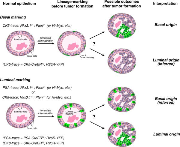
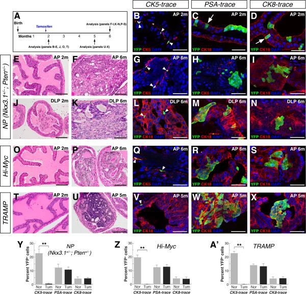
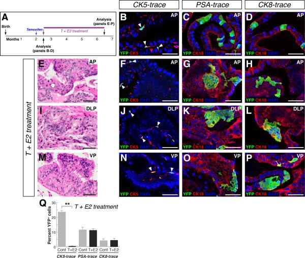
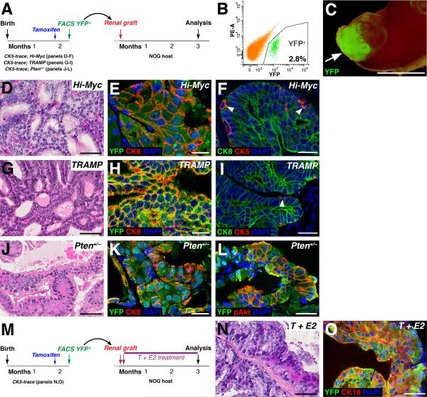
The identification of cell types of origin for cancer has important implications for tumor stratification and personalized treatment. For prostate cancer, the cell of origin has been intensively studied, but it has remained unclear whether basal or luminal epithelial cells, or both, represent cells of origin under physiological conditions in vivo. Here, we use a novel lineage-tracing strategy to assess the cell of origin in a diverse range of mouse models, including Nkx3.1(+/-); Pten(+/-), Pten(+/-), Hi-Myc, and TRAMP mice, as well as a hormonal carcinogenesis model. Our results show that luminal cells are consistently the observed cell of origin for each model in situ; however, explanted basal cells from these mice can generate tumors in grafts. Consequently, we propose that luminal cells are favored as cells of origin in many contexts, whereas basal cells only give rise to tumors after differentiation into luminal cells.


Figures

Figure 1
Figure 1. Experimental design for analysis of cell of origin
The inducible CK5-CreERT2 driver can lineage-mark basal cells by YFP expression in different prostate cancer models prior to overt cancer formation. Similarly, the inducible PSA-CreERT2 and CK8-CreERT2 drivers can mark luminal cells in phenotypically normal epithelium. The presence of YFP+ cell clusters in subsequent PIN/cancer lesions indicates that the marked cell type acts as the cell of origin in the mouse model analyzed.
Figure 2
Figure 2. Luminal cells are favored cells of origin in the Nkx3.1+/–; Pten+/– (NP), Hi-Myc, and TRAMP models
(A) Experimental time course. (B) Lineage-marking of basal cells (arrowheads) in the AP of CK5-trace; NP mice at 2 months of age. (C, D) Marking of luminal cells (arrows) in the AP of PSA-trace; NP mice (C) or CK8-trace mice (D) at 2 months. (E, F, J, K) H&E staining of NP prostates shows normal histology at 2 months, and PIN/carcinoma lesions at 6 months. (G, L) Clusters of YFP+ cells are rarely detected in CK5-trace; NP tumor lesions at 6 months. (H, I, M, N) YFP+ cell clusters in tumor lesions of PSA-trace; NP (H, M) and CK8-trace; NP (I, N) mice at 6 months. (O, P) Normal AP histology in Hi-Myc mice at 2 months (O), and PIN/carcinoma lesions at 6 months (P). (Q) Absence of YFP+ cell clusters in CK5-trace; Hi-Myc tumor lesions in the AP at 6 months. (R, S) YFP+ cell clusters in tumor lesions of PSA-trace; Hi-Myc mice (R) and CK8-trace; Hi-Myc mice (S) at 6 months. (T, U) Normal histology of the AP in TRAMP mice at 2 months (T), and carcinoma at 5 months (U). (V) Absence of YFP+ cell clusters in CK5-trace; TRAMP AP tumor lesions at 5 months. (W, X) YFP+ clusters in AP tumor lesions of PSA-trace; TRAMP (W) and CK8-trace; TRAMP (X) mice at 5 months. (Y-A’) Percentage of YFP+ cells in NP (Y), Hi-Myc (Z), and TRAMP (A’) models; Nor = normal, Tum = tumor; ** p<0.001 by Student's t-test; error bars are one standard deviation. Arrowheads in G, L, Q, V indicate marked basal cells. Scale bars in B-D, G-I, L-N, Q-S, and V-X correspond to 50 microns and in E, F, J, K, O, P, T, U to 100 microns. See also Figures S1-S4.
Figure 3
Figure 3. Luminal cells are the favored cell of origin of tumors induced by T+E2 hormonal treatment
(A) Experimental time course. (B-D) Lineage-marking of basal (arrowheads) and luminal cells in control CK5-trace (B), PSA-trace (C), and CK8-trace (D) mice. (E, I, M) PIN lesions in mice after T+E2 treatment. (F, J, N) YFP+ cell clusters are rarely detected in CK5-trace PIN lesions after T+E2 treatment; arrowheads indicate marked basal cells. (G, H, K, L, O, P) YFP+ clusters in PIN lesions of PSA-trace (G, K, O) and CK8-trace (H, L, P) mice after T+E2 treatment. (Q) Percentage of YFP+ cells; Cont = control untreated, T+E2 = treated; ** p<0.001 by Student's t-test; error bars are one standard deviation. Scale bars indicate 50 microns. See also Figures S2, S3.
Figure 4
Figure 4. Basal cells can give rise to prostate cancer in renal grafts
(A) Experimental design. (B) Representative flow-sort of YFP+ basal cells (2.8% of total prostate cells) from CK5-trace; Hi-Myc mice. (C) Kidney from recipient NOG mouse containing graft with YFP fluorescence (arrow). (D-I) Grafted basal cells from CK5-trace; Hi-Myc (D-F) or CK5-trace; TRAMP (G-I) mice generate PIN lesions (D, G), which contain mostly luminal cells (E, H) and some basal cells (arrowheads, F, I). (J-L) Basal cells from CK5-trace; Pten+/– mice generate PIN lesions (J) that contain mostly luminal cells (K) and express phospho-Akt (L). (M) Experimental design for T+E2 treatment of grafts. (N,O) Grafted basal cells give rise to PIN lesions (N) that contain mostly luminal cells (O) after T+E2 treatment. Scale bars in E, F, H, I, K correspond to 25 microns, in C to 5 mm, and in D, G, J, I, N, O to 50 microns.

References

    1. Blackwood JK, Williamson SC, Greaves LC, Wilson L, Rigas AC, Sandher R, Pickard RS, Robson CN, Turnbull DM, Taylor RW, et al. In situ lineage tracking of human prostatic epithelial stem cell fate reveals a common clonal origin for basal and luminal cells. J Pathol. 2011;225:181–188. - PubMed
    1. Blanpain C. Tracing the cellular origin of cancer. Nat Cell Biol. 2013;15:126–134. - PubMed
    1. Bosland MC, Ford H, Horton L. Induction at high incidence of ductal prostate adenocarcinomas in NBL/Cr and Sprague-Dawley Hsd:SD rats treated with a combination of testosterone and estradiol-17 beta or diethylstilbestrol. Carcinogenesis. 1995;16:1311–1317. - PubMed
    1. Choi N, Zhang B, Zhang L, Ittmann M, Xin L. Adult murine prostate basal and luminal cells are self-sustained lineages that can both serve as targets for prostate cancer initiation. Cancer Cell. 2012;21:253–265. - PMC - PubMed
    1. Ellwood-Yen K, Graeber TG, Wongvipat J, Iruela-Arispe ML, Zhang J, Matusik R, Thomas GV, Sawyers CL. Myc-driven murine prostate cancer shares molecular features with human prostate tumors. Cancer Cell. 2003;4:223–238. - PubMed

Publication types

MeSH terms