. 2014 Sep;23(133):308-19.
doi: 10.1183/09059180.00004914.

Differential diagnosis of usual interstitial pneumonia: when is it truly idiopathic?

Affiliations

Differential diagnosis of usual interstitial pneumonia: when is it truly idiopathic?

Wim A Wuyts et al. Eur Respir Rev. 2014 Sep.

Erratum in

  • Eur Respir Rev. 2014 Dec 23(134):537

Abstract

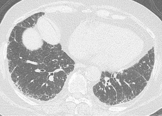
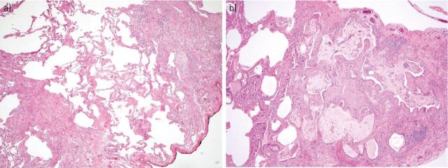
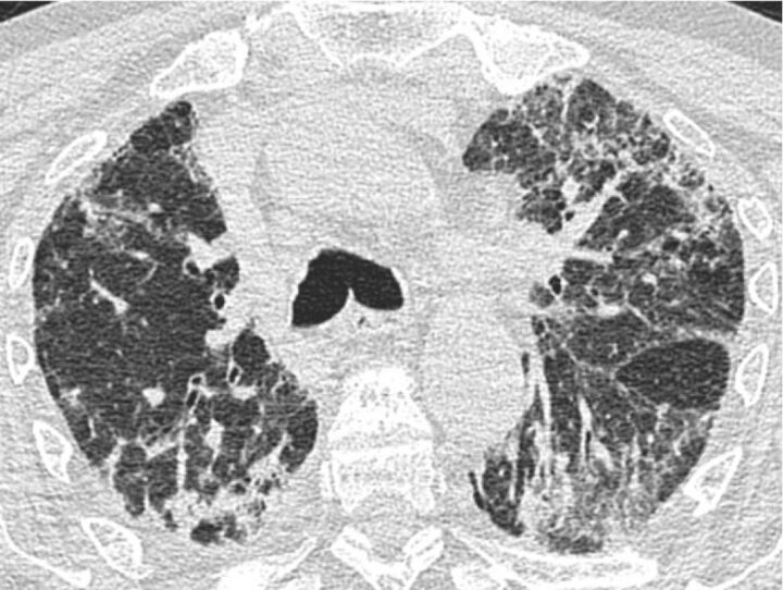
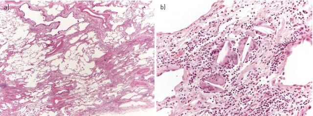
Idiopathic pulmonary fibrosis (IPF), the most common and lethal of the idiopathic interstitial pneumonias, is defined by a radiological and/or pathological pattern of usual interstitial pneumonia (UIP). However, UIP is not synonymous with IPF as other clinical conditions may be associated with UIP, including chronic hypersensitivity pneumonitis, collagen vascular disease, drug toxicity, asbestosis, familial IPF and Hermansky-Pudlak syndrome. Differentiating IPF ("idiopathic UIP") from conditions that mimic IPF ("secondary UIP") has substantial therapeutic and prognostic implications. A number of radiological and histological clues may help distinguish IPF from other conditions with a UIP pattern of fibrosis, but their appreciation requires extensive expertise in interstitial lung disease as well as an integrated multidisciplinary approach involving pulmonologists, radiologists and pathologists. In addition, multidisciplinary discussions may decrease the time to initial IPF diagnosis and, thus, enable more timely management. This concept was strongly emphasised by the 2011 ATS/ERS/JRS/ALAT guidelines. This article highlights, with the aid of a clinical case, the difficulties in making a diagnosis of IPF in clinical practice. Yet, an accurate diagnosis is critical, particularly given the availability of drugs that may reduce the pace of functional decline and disease progression in IPF.


Conflict of interest statement

Conflict of interest: Disclosures can be found alongside the online version of this article at err.ersjournals.com

Figures

Figure 1.
Figure 1.
A possible usual interstitial pneumonia pattern. Computed tomography scan showing subpleural and bibasal predominant reticular abnormality without honeycombing. Although this patient was highly likely to have idiopathic pulmonary fibrosis (in the appropriate clinical setting), surgical lung biopsy is needed to confirm the diagnosis.
Figure 2.
Figure 2.
a) Open lung biopsy showing fibrotic lung disease with heterogeneous spatial and temporal fibrosis consistent with a usual interstitial pneumonia pattern (haematoxylin and eosin stain, 40×). b) Honeycomb change with enlarged subpleural alveolar spaces with bronchiolar metaplasia, mucus plugs, and mild chronic inflammatory infiltrate (haematoxylin and eosin stain, 40×).
Figure 3.
Figure 3.
Chronic hypersensitivity pneumonitis with the computed tomography scan showing extensive reticular and ground-glass opacities. Traction bronchiectasis is also present in the apical segment of the left lower lobe. The left fissure is pulled apart because of the underlying fibrosis.
Figure 4.
Figure 4.
Usual interstitial pneumonia pattern in two patients with idiopathic pulmonary fibrosis. a) At low magnification, subpleural/paraseptal scars obscuring the alveolar architecture alternate with normal lung (“patchy fibrosis”). At the top, two areas of honeycombing can be observed (haematoxylin and eosin stain, 20×). b) At higher magnification a fibroblastic focus, consisting of a dome-shaped collection of fibroblasts/myofibroblasts embedded in a myxoid matrix and covered by hyperplastic pneumocytes, is observed. Note the pale grey colour of the fibroblastic focus, contrasting with the pink colour of the underlying scar (haematoxylin and eosin stain, 400×).
Figure 5.
Figure 5.
Chronic hypersensitivity pneumonitis with usual interstitial pneumonia pattern. a) At low magnification, bronchiolar damage with bridging fibrosis between bronchiole and pleura are the clues suggesting chronic hypersensitivity pneumonitis (haematoxylin and eosin stain, 20×). b) At higher magnification, a few inconspicuous interstitial granulomas, consisting of isolated giant cells with cholesterol clefts, can be observed (haematoxylin and eosin stain, 200×).

Comment in

References

    1. Raghu G, Collard HR, Egan JJ, et al. . An official ATS/ERS/JRS/ALAT statement: idiopathic pulmonary fibrosis: evidence-based guidelines for diagnosis and management. Am J Respir Crit Care Med 2011; 183: 788–824. - PMC - PubMed
    1. Noble PW, Barkauskas CE, Jiang D. Pulmonary fibrosis: patterns and perpetrators. J Clin Invest 2012; 122: 2756–2762. - PMC - PubMed
    1. Spagnolo P, Du Bois RM, Cottin V. Rare lung disease and orphan drug development. Lancet Respir Med 2013; 1: 479–487. - PubMed
    1. Spagnolo P, Tonelli R, Cocconcelli E, et al. . Idiopathic pulmonary fibrosis: diagnostic pitfalls and therapeutic challenges. Multidiscip Respir Med 2012; 7: 42. - PMC - PubMed
    1. Flaherty KR, Andrei AC, King TE, Jr, et al. . Idiopathic interstitial pneumonia: do community and academic physicians agree on diagnosis? Am J Respir Crit Care Med 2007; 175: 1054–1060. - PMC - PubMed

Publication types

MeSH terms

LinkOut - more resources