. 2014 Aug;18(4):307-11.
doi: 10.4196/kjpp.2014.18.4.307. Epub 2014 Aug 13.

Scopoletin from Cirsium setidens Increases Melanin Synthesis via CREB Phosphorylation in B16F10 Cells

Affiliations

Scopoletin from Cirsium setidens Increases Melanin Synthesis via CREB Phosphorylation in B16F10 Cells

Mi-Ja Ahn et al. Korean J Physiol Pharmacol. 2014 Aug.

Abstract

In this study, we isolated scopoletin from Cirsium setidens Nakai (Compositae) and tested its effects on melanogenesis. Scopoletin was not toxic to cells at concentrations less than 50 µM and increased melanin synthesis in a dose-dependent manner. As melanin synthesis increased, scopoletin stimulated the total tyrosinase activity, the rate-limiting enzyme of melanogenesis. In a cell-free system, however, scopoletin did not increase tyrosinase activity, indicating that scopoletin is not a direct activator of tyrosinase. Furthermore, Western blot analysis showed that scopoletin stimulated the production of microphthalmia-associated transcription factor (MITF) and tyrosinase expression via cAMP response element-binding protein (CREB) phosphorylation in a dose-dependent manner. Based on these results, preclinical and clinical studies are needed to assess the use of scopoletin for the treatment of vitiligo.

Keywords: CREB; Cirsium setidens; MITF; Scopoletin; Tyrosinase.


Figures

Fig. 1
Fig. 1
Structure of scopoletin.
Fig. 2
Fig. 2
Effect of scopoletin on B16F10 cell viability. B16F10 cells (2×10⁴ cells/well) were incubated for 24 h in serum-free DMEM, and then treated with 0.1, 1, 10, 20, or 50 µM scopoletin. After another 24 h, cell viability was determined by crystal violet assay. Results were expressed as % of the control. Each determination was made in triplicate and data shown represent the mean±S.D.
Fig. 3
Fig. 3
Effect of scopoletin on melanin content. B16F10 cells (5×10⁴ cells/well) were incubated with scopoletin (0.1, 1, 10, 20, or 50 µM) for 72 h. α-MSH (1 µM) was used as a positive control. Melanin released into the cell culture medium was measured as described in the materials and methods section. Each determination was made in triplicate and data shown represent the mean±S.D. *p<0.01 compared to the untreated control.
Fig. 4
Fig. 4
Effect of scopoletin on tyrosinase activity in B16F10 cells. B16F10 cells (5×10⁴ cells/well) were incubated with scopoletin (0.1, 1, 10, 20, or 50 µM) for 72 h. α-MSH (1 µM) was used as a positive control. Tyrosinase activity was measured as described in the materials and methods section. Each determination was made in triplicate and data shown represent the mean±S.D. *p<0.01 compared to the untreated control.
Fig. 5
Fig. 5
Effect of scopoletin on tyrosinase activity in a cell-free system. To test the direct effect of scopoletin on tyrosinase, tyrosinase activity was measured in a cell-free system as described in the materials and methods section. Each determination was made in triplicate and data shown represent the mean±S.D. *p<0.01 compared to the untreated control.
Fig. 6
Fig. 6
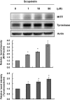
Effect of scopoletin on MITF and tyrosinase expression in B16F10 cells. Cells were exposed to 0~50 µM scopoletin for 72 h. MITF and tyrosinase expression were examined by Western blot analysis. Equal protein loading was confirmed by β-actin expression. Band intensity relative to control was determined by densitometric analysis. Values are expressed as mean±S.D. (n=3). *p<0.01 compared to untreated control.
Fig. 7
Fig. 7
Effect of scopoletin on the phosphorylation of CREB in B16F10 cells. Cells were treated with 1~50 µM scopoletin for 60 min (A), or exposed to 50 µM scopoletin for 0, 2, 10, 30, 60, 120, or 360 min (B). The phosphorylation of CREB was examined by Western blot analysis. Equal protein loading was confirmed by total-CREB expression. Band intensity relative to the control was determined by densitometric analysis. Values are expressed as mean±S.D. (n=3). *p<0.01 compared to untreated control.

References

    1. Hearing VJ, Tsukamoto K. Enzymatic control of pigmentation in mammals. FASEB J. 1991;5:2902–2909. - PubMed
    1. Cho YS, Lee KH, Park JW. Pyrithione-zinc Prevents UVB-induced Epidermal Hyperplasia by Inducing HIF-1alpha. Korean J Physiol Pharmacol. 2010;14:91–97. - PMC - PubMed
    1. Pichler R, Sfetsos K, Badics B, Gutenbrunner S, Auböck J. Vitiligo patients present lower plasma levels of alpha-melanotropin immunoreactivities. Neuropeptides. 2006;40:177–183. - PubMed
    1. Lee WB, Kwon HC, Cho OR, Lee KC, Choi SU, Baek NI, Lee KR. Phytochemical constituents of Cirsium setidens Nakai and their cytotoxicity against human cancer cell lines. Arch Pharm Res. 2002;25:628–635. - PubMed
    1. Tal B, Robeson DJ. The metabolism of sunflower phytoalexins ayapin and scopoletin: plant-fungus interactions. Plant Physiol. 1986;82:167–172. - PMC - PubMed