Review
. 2014 Aug 14:8:231.
doi: 10.3389/fncel.2014.00231. eCollection 2014.

Protection after stroke: cellular effectors of neurovascular unit integrity

Affiliations
Review

Protection after stroke: cellular effectors of neurovascular unit integrity

Rafael Andres Posada-Duque et al. Front Cell Neurosci. .

Abstract

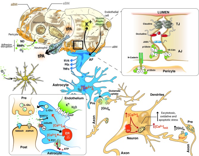
Neurological disorders are prevalent worldwide. Cerebrovascular diseases (CVDs), which account for 55% of all neurological diseases, are the leading cause of permanent disability, cognitive and motor disorders and dementia. Stroke affects the function and structure of blood-brain barrier, the loss of cerebral blood flow regulation, oxidative stress, inflammation and the loss of neural connections. Currently, no gold standard treatments are available outside the acute therapeutic window to improve outcome in stroke patients. Some promising candidate targets have been identified for the improvement of long-term recovery after stroke, such as Rho GTPases, cell adhesion proteins, kinases, and phosphatases. Previous studies by our lab indicated that Rho GTPases (Rac and RhoA) are involved in both tissue damage and survival, as these proteins are essential for the morphology and movement of neurons, astrocytes and endothelial cells, thus playing a critical role in the balance between cell survival and death. Treatment with a pharmacological inhibitor of RhoA/ROCK blocks the activation of the neurodegeneration cascade. In addition, Rac and synaptic adhesion proteins (p120 catenin and N-catenin) play critical roles in protection against cerebral infarction and in recovery by supporting the neurovascular unit and cytoskeletal remodeling activity to maintain the integrity of the brain parenchyma. Interestingly, neuroprotective agents, such as atorvastatin, and CDK5 silencing after cerebral ischemia and in a glutamate-induced excitotoxicity model may act on the same cellular effectors to recover neurovascular unit integrity. Therefore, future efforts must focus on individually targeting the structural and functional roles of each effector of neurovascular unit and the interactions in neural and non-neural cells in the post-ischemic brain and address how to promote the recovery or prevent the loss of homeostasis in the short, medium and long term.

Keywords: BBB; CDK5; NVU; Rho GTPases; p120 catenin; stroke.


Figures

Figure 1
Figure 1
NVU in cerebral ischemia includes a closely coupled signaling network of ECs, pericytes, astrocytes and neurons. Neurovascular uncoupling consists of the expression of various cytokines and adhesion molecules on ECs, promoting leukocyte adherence and accumulation, which thereby initiates an inflammatory response. Further, breakdown of the BBB via NO, MMPs and ROS permits neutrophil extravasation into the ischemic tissue in response to chemokines produced by astrocytes, macrophages, and microglia. Glutamate-mediated excitotoxicity via NMDA and AMPA receptors induces the hyperstimulation of neurons and subsequent calcium influx, indicating several intracellular calcium stores (mitochondria and ER), which triggers dendritotoxic and cell death pathways. Intrinsic neurovascular responses after stroke are triggered by astrocytes via the spread of calcium waves, promoting homeostatic gliotransmission. In addition, glutamate uptake by astrocytes, which requires EAATs, represents the primary neuroprotective response after excitotoxicity. Alternatively, K+ and glucose uptake and insulin and albumin transport signal the modulation of neurovascular homeostasis. Tight junctions (TJs) consisting of claudins, occludins and JAM and adherens junctions (AJ) including members of the cadherin-catenin system maintain the integrity of the endothelium. Cell–cell adhesion may transmit intracellular signals that regulate paracellular permeability, contact-induced inhibition of cell growth and new vessel formation. After stroke, pericytes display deformed junctions and increased formation of pinocytic vesicles, which affect the polarization of astrocyte end-feet (AF), detected as the defective deposition of the astrocyte-derived basement membrane (aBM). Deposition of the endothelium-derived basement membrane (eBM) is affected by the lack of pericytes. Moreover, modifications of the molecular organization and intracellular signaling of junction proteins may exert complex effects on vascular homeostasis.
Figure 2
Figure 2
Hypothetical model of the role of the effectors Rho GTPase, p120 catenin, and CDK5 in the recovery of the structure and function of the NVU after cerebral ischemia. CDK5 and RhoA/ROCK targeted to ECs, astrocytes and neurons may exert protection against excitotoxicity and promote neuronal plasticity and adhesion integrity. Reduction of CDK5 expression in neurons and astrocytes may facilitate synaptic plasticity, which requires NR2B for LTP and Rac1 and p120 catenin signaling for dendritic spine morphogenesis and synaptic adhesion. Decreased expression of CDK5 induces LTP via NR2B (GluN2B)-mediated excitatory postsynaptic currents, and the consequent induction or accumulation of p120ctn inhibits RhoA/ROCK, preventing F-actin retraction and inducing Rac1 activation, thus facilitating neurotransmission. In addition, CDK5 inhibition may prevent tau hyperphosphorylation and facilitate glutamate uptake in astrocytes, preserve actin remodeling and promote angiogenesis via eNOS activation. In turn, ROCK inhibition also prevents tau hyperphosphorylation and dendritic spine retraction and increases the expression of EAATs to enhance glutamate uptake and promote BBB integrity. Moreover, actin remodeling induced by Tiam/Rac1 favors stellation and survival in astrocytes. Likewise, the signaling molecules VE-cadherin and p120 catenin activate Rac/PI3 kinase and suppress RhoA to maintain the survival and organization of the actin cytoskeleton, form complexes with VEGFR-2 to promote BBB adhesion and integrity, and prevent neutrophil diapedesis. ROCK and CDK5 targeting may promote calcium wave stimulation via glutamate receptor and PLC/IP3 signaling in astrocytes to facilitate NO and PGE2 release, thus regulating endothelial vasodilatation and vasoconstriction.

References

    1. Abbott N. J. (2000). Inflammatory mediators and modulation of blood-brain barrier permeability. Cell. Mol. Neurobiol. 20, 131–147 10.1023/A:1007074420772 - DOI - PMC - PubMed
    1. Abbott N. J., Ronnback L., Hansson E. (2006). Astrocyte-endothelial interactions at the blood-brain barrier. Nat. Rev. Neurosci. 7, 41–53 10.1038/nrn1824 - DOI - PubMed
    1. Adelson J. D., Barreto G. E., Xu L., Kim T., Brott B. K., Ouyang Y. B., et al. (2012). Neuroprotection from stroke in the absence of MHCI or PirB. Neuron 73, 1100–1107 10.1016/j.neuron.2012.01.020 - DOI - PMC - PubMed
    1. Aghajanian A., Wittchen E. S., Allingham M. J., Garrett T. A., Burridge K. (2008). Endothelial cell junctions and the regulation of vascular permeability and leukocyte transmigration. J. Thromb. Haemost. 6, 1453–1460 10.1111/j.1538-7836.2008.03087.x - DOI - PMC - PubMed
    1. Alavi A., Clark C., Fazekas F. (1998). Cerebral ischemia and Alzheimer’s disease: critical role of PET and implications for therapeutic intervention. J. Nucl. Med. 39, 1363–1365 - PubMed

LinkOut - more resources