. 2014 Aug 15:8:81.
doi: 10.3389/fnana.2014.00081. eCollection 2014.

Towards a unified scheme of cortical lamination for primary visual cortex across primates: insights from NeuN and VGLUT2 immunoreactivity

Affiliations

Towards a unified scheme of cortical lamination for primary visual cortex across primates: insights from NeuN and VGLUT2 immunoreactivity

Pooja Balaram et al. Front Neuroanat. .

Abstract

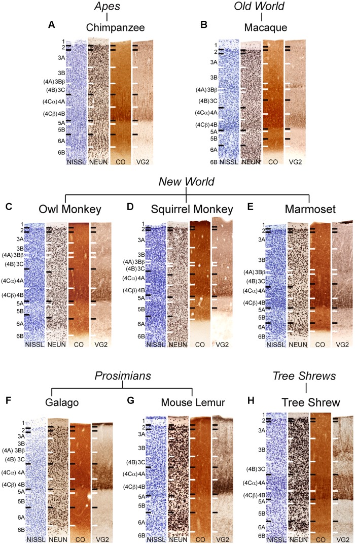
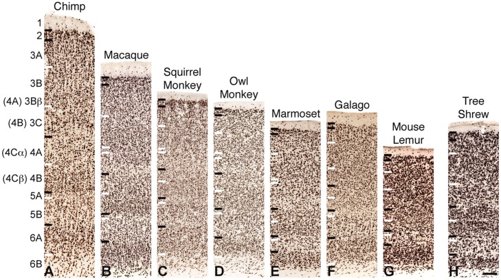
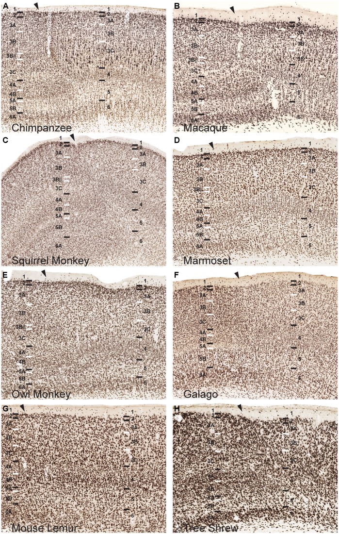
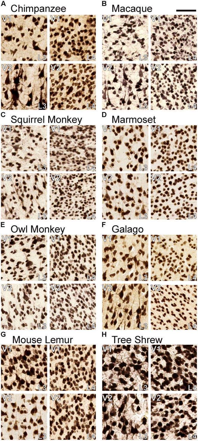
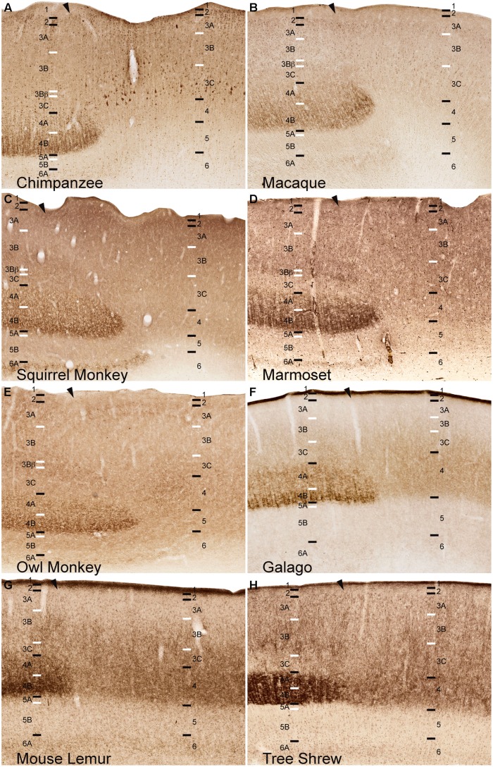
Primary visual cortex (V1) is clearly distinguishable from other cortical areas by its distinctive pattern of neocortical lamination across mammalian species. In some mammals, primates in particular, the layers of V1 are further divided into a number of sublayers based on their anatomical and functional characteristics. While these sublayers are easily recognizable across a range of primates, the exact number of divisions in each layer and their relative position within the depth of V1 has been inconsistently reported, largely due to conflicting schemes of nomenclature for the V1 layers. This conflict centers on the definition of layer 4 in primate V1, and the subdivisions of layer 4 that can be consistently identified across primate species. Brodmann's (1909) laminar scheme for V1 delineates three subdivisions of layer 4 in primates, based on cellular morphology and geniculate inputs in anthropoid monkeys. In contrast, Hässler's (1967) laminar scheme delineates a single layer 4 and multiple subdivisions of layer 3, based on comparisons of V1 lamination across the primate lineage. In order to clarify laminar divisions in primate visual cortex, we performed NeuN and VGLUT2 immunohistochemistry in V1 of chimpanzees, Old World macaque monkeys, New World squirrel, owl, and marmoset monkeys, prosimian galagos and mouse lemurs, and non-primate, but highly visual, tree shrews. By comparing the laminar divisions identified by each method across species, we find that Hässler's (1967) laminar scheme for V1 provides a more consistent representation of neocortical layers across all primates, including humans, and facilitates comparisons of V1 lamination with non-primate species. These findings, along with many others, support the consistent use of Hässler's laminar scheme in V1 research.

Keywords: Brodmann; Hässler; NeuN; cortical layers; primate; tree shrew; visual cortex.


Figures

FIGURE 1
FIGURE 1
Laminar distributions of Nissl, neuronal nuclear antigen (NeuN), cytochrome oxidase (CO), and vesicular glutamate transporter 2 (VGLUT2) in primary visual cortex of chimpanzees (A), Old World macaque monkeys (B), owl monkeys (C), New World squirrel monkeys (D), and marmosets (E), prosimian galagos (F) and mouse lemurs (G), and tree shrews (H). Hässler’s (1967) laminar divisions listed to the left, Brodmann’s (1909) divisions are listed in parentheses. Individual panels are scaled relative to V1 of chimpanzees to visualize comparisons between laminar density and staining intensity across primate species. Absolute scale comparisons are shown in Figure 2.
FIGURE 2
FIGURE 2
NeuN immunoreactivity through the cortical layers of V1 in (A) chimpanzees, (B) macaque monkeys, (C) squirrel monkeys, (D) owl monkeys, (E) marmosets, (F) galagos, (G) mouse lemurs, and (H) tree shrews. Laminar designations by Hässler (1967) are listed to the left, Brodmann’s divisions are listed in parentheses. Black lines indicate neocortical layer boundaries and white lines indicate sublaminar boundaries within each layer. Scale bar is 250um.
FIGURE 3
FIGURE 3
NeuN immunoreactivity reveals transitions of laminar boundaries between V1 and V2 in (A) chimpanzees, (B) macaque monkeys, (C) squirrel monkeys, (D) marmosets, (E) owl monkeys, (F) galagos, (G) mouse lemurs, and (H) tree shrews. Hässler’s laminar designations are listed on each panel for both visual areas. Arrowheads demarcate the V1/V2 border, V1 is to the left of the arrowhead and V2 is to the right of the arrowhead in each panel. Black lines indicate laminar boundaries and white lines indicate sublaminar boundaries in each area. Individual panels are scaled relative to V1 of chimpanzees to visualize laminar transitions between V1 and V2 across species.
FIGURE 4
FIGURE 4
Comparisons of neuronal density in layers 3C and 4 of (A) chimpanzees, (B) macaque monkeys, (C) squirrel monkeys, (D) marmosets, (E) owl monkeys, (F) galagos, (G) mouse lemurs, and (H) tree shrews. In both areas, neurons in layer 4 are significantly more densely packed than neurons in layer 3C. Scale bar is 20 μm.
FIGURE 5
FIGURE 5
VGLUT2 immunoreactivity reveals the continuation of layer 4, but not layer 3Bβ, at the boundary of V1 into V2 across (A) chimpanzees, (B) macaque monkeys, (C) squirrel monkeys, (D) marmosets, (E) owl monkeys, (F) galagos, (G) mouse lemurs, and (H) tree shrews. Hässler’s laminar designations are listed on each panel for both visual areas. Arrowheads demarcate the V1/V2 border, V1 is to the left of the arrowhead and V2 is to the right of the arrowhead in each panel. Black lines indicate laminar boundaries and white lines indicate sublaminar boundaries in each area. Individual panels are scaled relative to V1 of chimpanzees to visualize laminar transitions between V1 and V2 across species.

References

    1. Aihara Y., Mashima H., Onda H., Hisano S., Kasuya H., Hori T., et al. (2000). Molecular cloning of a novel brain-type Na(+)-dependent inorganic phosphate cotransporter. J. Neurochem. 74 2622–2625 10.1046/j.1471-4159.2000.0742622.x - DOI - PubMed
    1. Allman J. M., Kaas J. H. (1971). Representation of the visual field in striate and adjoining cortex of the owl monkey (Aotus trivirgatus). Brain Res. 35 89–106 10.1016/0006-8993(71)90596-8 - DOI - PubMed
    1. Allman J., McGuinness E. (1988). “Visual cortex in primates,” in Neurosciences: Comparative Primate Biology eds Steklis H. D., Erwi J. (New York, NY: Alan R. Liss Inc.) 279–326
    1. Balaram P., Hackett T. A., Kaas J. H. (2013). Differential expression of vesicular glutamate transporters 1 and 2 may identify distinct modes of glutamatergic transmission in the macaque visual system. J. Chem. Neuroanat. 50–51, 21–38 10.1016/j.jchemneu.2013.02.007 - DOI - PMC - PubMed
    1. Balaram P., Takahata T., Kaas J. H. (2011). VGLUT2 mRNA and protein expression in the visual thalamus and midbrain of prosimian galagos (Otolemur garnetti). Eye Brain 2011 5–15 10.2147/EB.S16998 - DOI - PMC - PubMed

LinkOut - more resources