Review
. 2014 Aug 15:5:307.
doi: 10.3389/fphys.2014.00307. eCollection 2014.

Peroxisome biogenesis in mammalian cells

Affiliations
Review

Peroxisome biogenesis in mammalian cells

Yukio Fujiki et al. Front Physiol. .

Abstract

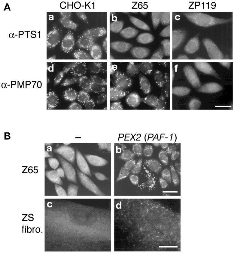
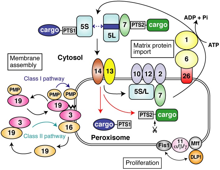
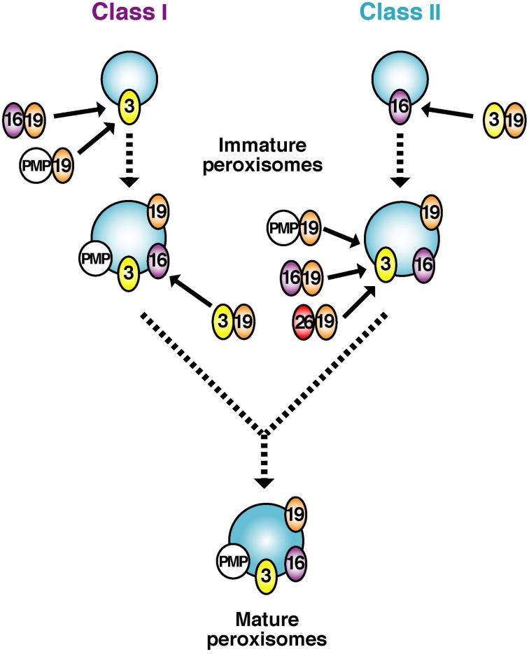
To investigate peroxisome assembly and human peroxisome biogenesis disorders (PBDs) such as Zellweger syndrome, thirteen different complementation groups (CGs) of Chinese hamster ovary (CHO) cell mutants defective in peroxisome biogenesis have been isolated and established as a model research system. Successful gene-cloning studies by a forward genetic approach utilized a rapid functional complementation assay of CHO cell mutants led to isolation of human peroxin (PEX) genes. Search for pathogenic genes responsible for PBDs of all 14 CGs is now completed together with the homology search by screening the human expressed sequence tag database using yeast PEX genes. Peroxins are divided into three groups: (1) peroxins including Pex3p, Pex16p, and Pex19p, are responsible for peroxisome membrane biogenesis via classes I and II pathways; (2) peroxins that function in matrix protein import; (3) those such as three forms of Pex11p, Pex11pα, Pex11pβ, and Pex11pγ, are involved in peroxisome proliferation where DLP1, Mff, and Fis1 coordinately function. In membrane assembly, Pex19p forms complexes in the cytosol with newly synthesized PMPs including Pex16p and transports them to the receptor Pex3p, whereby peroxisomal membrane is formed (Class I pathway). Pex19p likewise forms a complex with newly made Pex3p and translocates it to the Pex3p receptor, Pex16p (Class II pathway). In matrix protein import, newly synthesized proteins harboring peroxisome targeting signal type 1 or 2 are recognized by Pex5p or Pex7p in the cytoplasm and are imported to peroxisomes via translocation machinery. In regard to peroxisome-cytoplasmic shuttling of Pex5p, Pex5p initially targets to an 800-kDa docking complex consisting of Pex14p and Pex13p and then translocates to a 500-kDa RING translocation complex. At the terminal step, Pex1p and Pex6p of the AAA family mediate the export of Pex5p, where Cys-ubiquitination of Pex5p is essential for the Pex5p exit.

Keywords: CHO cell mutants; Zellweger syndrome; genetic phenotype-complementation; import machinery; membrane assembly; pathogenic genes; peroxins; peroxisome targeting signals.


Figures

Figure 1
Figure 1
Morphology of peroxisomes in CHO cell mutants defective in peroxisome biogenesis and cloning pathogenic genes of PBDs. (A) Cells are stained with antibodies to PTS1 (a–c) and PMP70 (d–f). Cells are as indicated at the top. Scale bar, 20 μm. In contrast to the wild-type CHO-K1 cells, PTS1 proteins are discernible in the cytosol in pex2 Z65 and pex19 ZP119. Z65 contains PMP70-positive peroxisomal remnants, whilst ZP119 is absent from such peroxisome ghosts, indicative of the defect of membrane protein import. (B) Cloning of pathogenic gene of PBD. Peroxisome-restoring PEX gene were isolated by functional complementation assay using CHO mutant. Restoration of peroxisomes in Z65 (a) by transfection of rat liver cDNA library (b). Transformed cells positive in catalase import contained PAF-1 (PEX2). In fibroblasts from a patient with ZS of CG10 (c), expression of PAF-1 restored the impaired import of catalase (d). Scale bar, 20 μm (a,b); 30 μm (c,d).
Figure 2
Figure 2
A schematic view of peroxisome biogenesis in mammalian cells. The subcellular localization and molecular characteristics of peroxins are shown. Peroxins are classified into three groups: (1) peroxins that are required for matrix protein import; (2) those including Pex3p, Pex16p and Pex19p, responsible for peroxisome membrane assembly via classes I and II pathways (see in this figure); (3) those such as three forms of Pex11p, Pex11pα, Pex11pβ, and Pex11pγ, apparently involved in peroxisome proliferation where DLP1, Mff, and Fis1 coordinately function. PTS1 and PTS2 proteins are recognized by Pex5p and Pex7p, respectively, in the cytoplasm. Two isoforms, Pex5pS and Pex5pL, of Pex5p are identified in mammals. PTS1 proteins are transported by homo- and hetero-oligomers of Pex5pS and Pex5pL to peroxisomes, where Pex14p functions as a convergent, initial docking site of the “protein import machinery” translocon. Pex5pL directly interacts with the PTS2 receptor, Pex7p, carrying its cargo PTS2 protein in the cytosol and translocates the Pex7p–PTS2 protein complex to Pex14p. PTS1 and PTS2 proteins are then released at the inner surface and/or inside of peroxisomes, downstream Pex14p and upstream Pex13p. Pex5p and Pex7p subsequently translocate to other translocon components, named translocation complex comprising the RING peroxins, Pex2p, Pex10p, and Pex12p. Both Pex5p and Pex7p finally shuttle back to the cytosol. In regard to peroxisome-cytoplasmic shuttling of Pex5p, Pex5p initially targets to an 800-kDa docking complex containing Pex14p and then translocates to a 500-kDa translocation complex comprising RING peroxins. At the terminal step of the protein import reaction, Pex1p and Pex6p of the AAA family catalyze the export of Pex5p, where Cys-ubiquitination of Pex5p is prerequisite to the Pex5p exit.
Figure 3
Figure 3
A model for early stages of peroxisomal membrane biogenesis involving mutually dependent targeting of Pex3p and Pex16p, named classes I and II pathways. The initial membranes harboring Pex3p or Pex16p culminate in indistinguishable, matured peroxisomes. Pex19p forms complexes in the cytosol with newly synthesized PMPs including Pex16p and C-tailed anchored membrane proteins such as Pex26p and transports them to the membrane protein receptor Pex3p, whereby peroxisome membrane is assembled (Class I pathway). With respect to biogenesis of Pex3p, Pex19p likewise forms a complex with newly synthesized Pex3p and translocates it to the Pex3p receptor, Pex16p (Class II pathway). Of note, peroxisomes are assembled no matter which pathway initially proceeds.

References

    1. Abe I., Fujiki Y. (1998). cDNA cloning and characterization of a constitutively expressed isoform of the human peroxin Pex11p. Biochem. Biophys. Res. Commun. 252, 529–533 10.1006/bbrc.1998.9684 - DOI - PubMed
    1. Abe I., Okumoto K., Tamura S., Fujiki Y. (1998). Clofibrate-inducible, 28-kDa peroxisomal integral membrane protein is encoded by PEX11. FEBS Lett. 431, 468–472 10.1016/S0014-5793(98)00815-1 - DOI - PubMed
    1. Agrawal G., Joshi S., Subramani S. (2011). Cell-free sorting of peroxisomal membrane proteins from the endoplasmic reticulum. Proc. Natl. Acad. Sci. U.S.A. 108, 9113–9118 10.1073/pnas.1018749108 - DOI - PMC - PubMed
    1. Baerends R. J. S., Rasmussen S. W., Hilbrands R. E., Van Der Heide M., Faber K. N., Reuvekamp P. T. W., et al. (1996). The Hansenula polymorpha PER9 gene encodes a peroxisomal membrane protein essential for peroxisome assembly and integrity. J. Biol. Chem. 271, 8887–8894 10.1074/jbc.271.15.8887 - DOI - PubMed
    1. Carvalho A. F., Pinto M. P., Grou C. P., Alencastre I. S., Fransen M., Sá-Miranda C., et al. (2007). Ubiquitination of mammalian Pex5p, the peroxisomal import receptor. J. Biol. Chem. 282, 31267–31272 10.1074/jbc.M706325200 - DOI - PubMed