

Evaluation of implant loosening following segmental pedicle screw fixation in adolescent idiopathic scoliosis: a 2 year follow-up with low-dose CT
- PMID: 25177357
- PMCID: PMC4149778
- DOI: 10.1186/1748-7161-9-13
Evaluation of implant loosening following segmental pedicle screw fixation in adolescent idiopathic scoliosis: a 2 year follow-up with low-dose CT
Abstract
Background: The long term radiological status of screw fixation following scoliosis surgery with all pedicle screw construct is not previously studied.
Aim: To evaluate the incidence of loosening (implant failure) evaluated with low-dose CT two years following scoliosis surgery.
Study design: Retrospective study.
Methods: 81 consecutive patients with adolescent idiopathic scoliosis (AIS), aged 18 ± 3 years at 2 years follow-up (83% were female), subjected for scoliosis corrective surgery with all pedicle screw construct (total of 1666 screws) has been examined with plain radiography and with low dose CT 6 weeks and 2 years postoperatively.
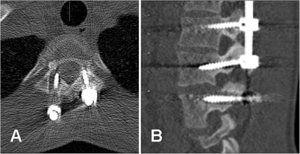
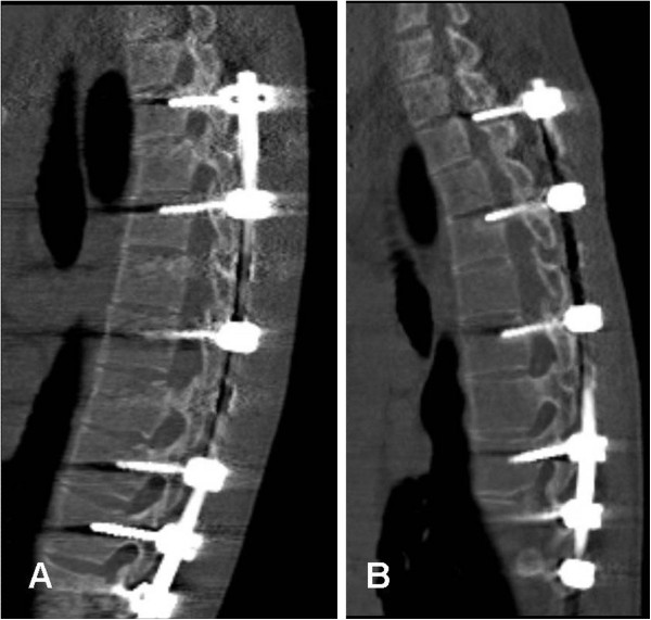
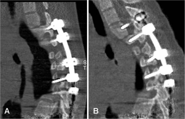
Results: In 26 out of 81 (32%) patients there were signs of loosening of one or more screws, a maximum 3 screws. 47 out of 1666 (2.8%) screws showed evidence of loosening. Preoperative Cobb angle was 56° among patients with loosening compared with 53° among patients with no evidence of loosening (P = 0.288). In males there were signs of loosening in 8 out of 14 (57%) and in females 18 out of 67 (27%), (P = 0.027). Among cases with loosening, 14% had suboptimal screw placement at the first postoperative CT compared with 11% among patients with no evidence of loosening (P = 0.254). One patient with a loosened L4 screw had neurological deficit and subjected for revision of the construct. Out of 26 patients with evidence of loosening, 5 patients reported minor pain or discomfort, 1 patient had a minor proximal junctional kyphosis of about 15° and 3 patients showed evidence of pull-out of 3-5 mm at the upper end of the construct but no clinical complaint. With plain radiography loosening could be observed only in 11 out of 26 cases, 5 were in the lumbar region.
Conclusions: In a consecutive series of 81 cases with AIS who had underwent scoliosis surgery, one third showed, 2 years after the intervention, minor screw loosening. Males were more prone to develop screw loosening. In CT system that enables low-dose protocol, CT is recommended for the evaluation of evidence of screw loosening.
Figures
References
-
- Abul-Kasim K, Overgaard A, Maly P, Ohlin A, Gunnarsson M, Sundgren PC. Low-dose helical computed tomography (CT) in the perioperative workup of adolescent idiopathic scoliosis. Eur Radiol. 2009;19(3):610–618. - PubMed
-
- Abul-Kasim K, Strombeck A, Ohlin A, Maly P, Sundgren PC. Reliability of low-radiation dose CT in the assessment of screw placement after posterior scoliosis surgery, evaluated with a new grading system. Spine. 2009;34(9):941–948. - PubMed
-
- Ohtori S, Inoue G, Orita S, Yamauchi K, Eguchi Y, Ochiai N, Kishida S, Kuniyoshi K, Aoki Y, Nakamura J, Ishikawa T, Miyagi M, Kamoda H, Suzuki M, Kubota G, Sakuma Y, Oikawa Y, Inage K, Sainoh T, Takaso M, Toyone T, Takahashi K. Comparison of Teriparatide and Bisphosphonate Treatment to Reduce Pedicle Screw Loosening After Lumbar Spinal Fusion Surgery in Postmenopausal Women with Osteoporosis from a Bone Quality Perspective. Spine. 2013;38(8):E487–492. - PubMed
-
- Wu ZX, Gong FT, Liu L, Ma ZS, Zhang Y, Zhao X, Yang M, Lei W, Sang HX. A comparative study on screw loosening in osteoporotic lumbar spine fusion between expandable and conventional pedicle screws. Arch Orthop Trauma Surg. 2012;132(4):471–476. 10.1007/s00402-011-1439-6. - PubMed
LinkOut - more resources
Full Text Sources
Other Literature Sources
