

A Study on the Nature of Association between Demodex Mites and Bacteria Involved in Skin and Meibomian Gland Lesions of Demodectic Mange in Cattle
- PMID: 25177514
- PMCID: PMC4142185
- DOI: 10.1155/2014/413719
A Study on the Nature of Association between Demodex Mites and Bacteria Involved in Skin and Meibomian Gland Lesions of Demodectic Mange in Cattle
Abstract
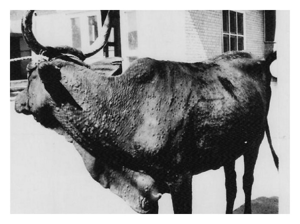
The nature of association between Demodex mites and bacteria involved in bovine demodectic mange lesions and the normal flora inhabiting the skin of noninfected animals was investigated. Demodex bovis and D. ghanensis mites were isolated from the infected purulent material extracted from skin and meibomian gland lesions, respectively. The mites could not be demonstrated in skin brushings or impression smears from the eyes of noninfected cattle. Pathogenic bacteria (Staphylococcus aureus and Streptococcus pyogenes (Group A)) and opportunistic organisms (Proteus vulgaris, Pseudomonas aeruginosa, Staphylococcus epidermidis, and Trueperella pyogenes) were isolated from skin lesions of demodectic mange, and Moraxella bovis and Staphylococcus aureus were isolated from meibomian gland lesions. Bacillus subtilis, Escherichia coli, Proteus vulgaris, Staphylococcus aureus, Staphylococcus epidermidis, and Streptococcus pyogenes (Group A) were isolated from skin brushings from noninfected cattle. The nature of association between Demodex mites and bacteria in demodectic mange lesions is synergistic and of equal significance. Pathogenic and opportunistic bacteria facilitated the establishment of Demodex mites in the lesions produced and provided an excellent microclimate for the mites to propagate and reproduce, resulting in severe and progressive disease. The "high-turnover" granulomatous reaction which characterized the histopathological changes proved that Demodex mites and associated bacteria were persistent and immunogenic.
Figures


Similar articles
-
Under the lash: Demodex mites in human diseases.Biochem (Lond). 2009 Aug 1;31(4):2-6. Biochem (Lond). 2009. PMID: 20664811 Free PMC article.
-
[Demodex folliculorum and rosacea: experimental and immunological studies].Z Hautkr. 1980 Sep 15;55(18):1211-8. Z Hautkr. 1980. PMID: 6451097 German.
-
Demodex mites: facts and controversies.Clin Dermatol. 2010 Sep-Oct;28(5):502-4. doi: 10.1016/j.clindermatol.2010.03.006. Clin Dermatol. 2010. PMID: 20797509
-
Potential role of Demodex mites and bacteria in the induction of rosacea.J Med Microbiol. 2012 Nov;61(Pt 11):1504-1510. doi: 10.1099/jmm.0.048090-0. Epub 2012 Aug 29. J Med Microbiol. 2012. PMID: 22933353 Review.
-
Demodex mites.Clin Dermatol. 2014 Nov-Dec;32(6):739-43. doi: 10.1016/j.clindermatol.2014.02.012. Epub 2014 Feb 28. Clin Dermatol. 2014. PMID: 25441466 Review.
Cited by
-
Genomic and anatomical comparisons of skin support independent adaptation to life in water by cetaceans and hippos.Curr Biol. 2021 May 24;31(10):2124-2139.e3. doi: 10.1016/j.cub.2021.02.057. Epub 2021 Apr 1. Curr Biol. 2021. PMID: 33798433 Free PMC article.
-
Nasopulmonary mites (Acari: Halarachnidae) as potential vectors of bacterial pathogens, including Streptococcus phocae, in marine mammals.PLoS One. 2022 Jun 16;17(6):e0270009. doi: 10.1371/journal.pone.0270009. eCollection 2022. PLoS One. 2022. PMID: 35709209 Free PMC article.
-
Significance and Roles of Proteus spp. Bacteria in Natural Environments.Microb Ecol. 2016 Nov;72(4):741-758. doi: 10.1007/s00248-015-0720-6. Epub 2016 Jan 9. Microb Ecol. 2016. PMID: 26748500 Free PMC article. Review.
-
Comparison of the efficacy of tea tree (Melaleuca alternifolia) oil with other current pharmacological management in human demodicosis: A Systematic Review.Parasitology. 2020 Dec;147(14):1587-1613. doi: 10.1017/S003118202000150X. Epub 2020 Aug 10. Parasitology. 2020. PMID: 32772960 Free PMC article.
References
-
- Kaufmann J. Parasitic Infections of Domestic Animals: A Diagnostic Manual. Berlin, Germany: Birkhäuser; 1996.
-
- OIE. Mange. Terrestrial Manual, Chapter 2. 9. 8., 2013.
-
- Scott DW. Color Atlas of Farm Animal Dermatology. 1st edition. Blackwell Australia Publishing; 2007.
-
- Soulsby EJL. Helminths , Arthropods and Protozoa of Domesicated Animals. 7th edition. London, UK: Bailliere Tindall; 1982.
-
- Urquhart GM, Armour J, Duncan JL, Dunn AM, Jennings FW. Veterinary Parasitology. 2nd edition. Blackwell; 2010.
LinkOut - more resources
Full Text Sources
Other Literature Sources
