

Endogenous Tetrapyrroles Influence Leukocyte Responses to Lipopolysaccharide in Human Blood: Pre-Clinical Evidence Demonstrating the Anti-Inflammatory Potential of Biliverdin
- PMID: 25177524
- PMCID: PMC4145741
- DOI: 10.4172/2155-9899.1000218
Endogenous Tetrapyrroles Influence Leukocyte Responses to Lipopolysaccharide in Human Blood: Pre-Clinical Evidence Demonstrating the Anti-Inflammatory Potential of Biliverdin
Abstract
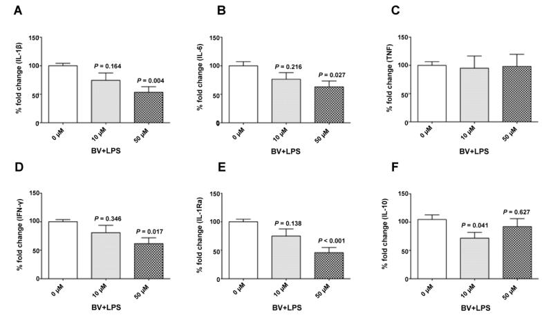
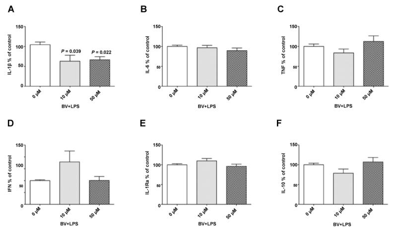
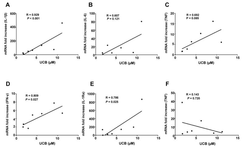
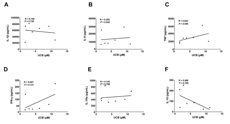
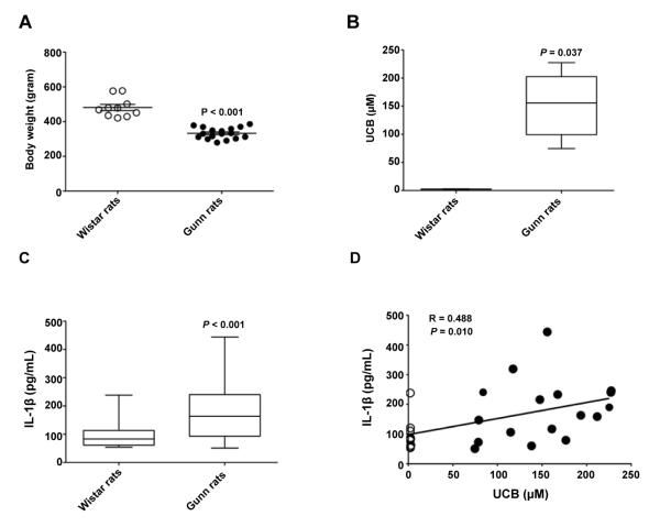
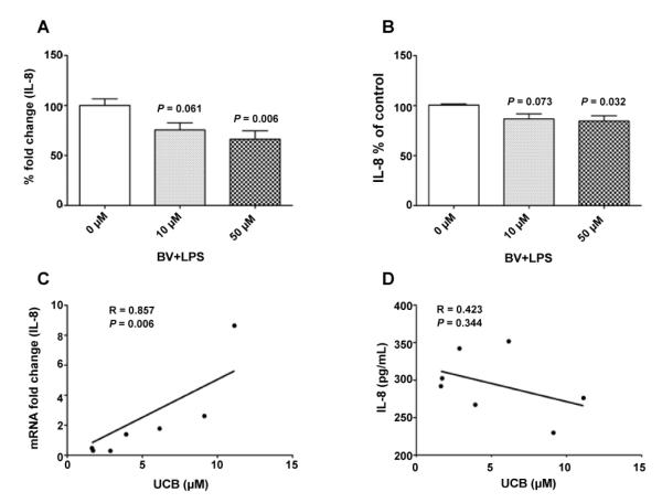
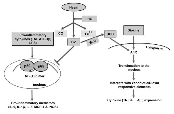
Sepsis is associated with abnormal host immune function in response to pathogen exposure, including endotoxin (lipopolysaccharide; LPS). Cytokines play crucial roles in the induction and resolution of inflammation in sepsis. Therefore, the primary aim of this study was to investigate the effects of endogenous tetrapyrroles, including biliverdin (BV) and unconjugated bilirubin (UCB) on LPS-induced cytokines in human blood. Biliverdin and UCB are by products of haem catabolism and have strong cytoprotective, antioxidant and anti-inflammatory effects. In the present study, whole human blood supplemented with BV and without was incubated in the presence or absence of LPS for 4 and 8 hours. Thereafter, whole blood was analysed for gene and protein expression of cytokines, including IL-1β, IL-6, TNF, IFN-γ, IL-1Ra and IL-8. Biliverdin (50 μM) significantly decreased the LPS-mediated gene expression of IL-1β, IL-6, IFN-γ, IL-1Ra and IL-8 (P<0.05). Furthermore, BV significantly decreased LPS-induced secretion of IL-1β and IL-8 (P<0.05). Serum samples from human subjects and, wild type and hyperbilirubinaemic Gunn rats were also used to assess the relationship between circulating bilirubin and cytokine expression/production. Significant positive correlations between baseline UCB concentrations in human blood and LPS-mediated gene expression of IL-1β (R=0.929), IFN-γ (R=0.809), IL-1Ra (R=0.786) and IL-8 (R=0.857) were observed in blood samples (all P<0.05). These data were supported by increased baseline IL-1β concentrations in hyperbilirubinaemic Gunn rats (P<0.05). Blood samples were also investigated for complement receptor-5 (C5aR) expression. Stimulation of blood with LPS decreased gene expression of C5aR (P<0.05). Treatment of blood with BV alone and in the presence of LPS tended to decrease C5aR expression (P=0.08). These data indicate that supplemented BV inhibits the ex vivo response of human blood to LPS. Surprisingly, however, baseline UCB was associated with heighted inflammatory response to LPS. This is the first study to explore the effects of BV in a preclinical human model of inflammation and suggests that BV could represent an anti-inflammatory target for the prevention of LPS mediated inflammation in vivo.
Keywords: Cytokine; Inflammation; Lipopolysaccharide; Tetrapyrroles.
Figures
Similar articles
-
Biliverdin and bilirubin sulfonate inhibit monosodium urate induced sterile inflammation in the rat.Eur J Pharm Sci. 2020 Dec 1;155:105546. doi: 10.1016/j.ejps.2020.105546. Epub 2020 Sep 12. Eur J Pharm Sci. 2020. PMID: 32927072
-
Biliverdin modulates the expression of C5aR in response to endotoxin in part via mTOR signaling.Biochem Biophys Res Commun. 2014 Jun 20;449(1):94-9. doi: 10.1016/j.bbrc.2014.04.150. Epub 2014 May 9. Biochem Biophys Res Commun. 2014. PMID: 24814708 Free PMC article.
-
Role of endogenous interleukin-1 receptor antagonist in regulating fever induced by localised inflammation in the rat.J Physiol. 2001 Feb 15;531(Pt 1):171-80. doi: 10.1111/j.1469-7793.2001.0171j.x. J Physiol. 2001. PMID: 11179401 Free PMC article.
-
Role of several mediators of inflammation on the mouse hypothalamo-pituitary-adrenal axis response during acute endotoxemia.Neuroimmunomodulation. 1999 Sep-Oct;6(5):336-43. doi: 10.1159/000026393. Neuroimmunomodulation. 1999. PMID: 10474052
-
Thymoquinone increases the expression of neuroprotective proteins while decreasing the expression of pro-inflammatory cytokines and the gene expression NFκB pathway signaling targets in LPS/IFNγ -activated BV-2 microglia cells.J Neuroimmunol. 2018 Jul 15;320:87-97. doi: 10.1016/j.jneuroim.2018.04.018. Epub 2018 May 4. J Neuroimmunol. 2018. PMID: 29759145 Free PMC article.
Cited by
-
Heme Oxygenases in Cardiovascular Health and Disease.Physiol Rev. 2016 Oct;96(4):1449-508. doi: 10.1152/physrev.00003.2016. Physiol Rev. 2016. PMID: 27604527 Free PMC article. Review.
-
Reduced Biliverdin Reductase-A Expression in Visceral Adipose Tissue is Associated with Adipocyte Dysfunction and NAFLD in Human Obesity.Int J Mol Sci. 2020 Nov 29;21(23):9091. doi: 10.3390/ijms21239091. Int J Mol Sci. 2020. PMID: 33260451 Free PMC article.
-
Bilirubin Attenuates ER Stress-Mediated Inflammation, Escalates Apoptosis and Reduces Proliferation in the LS174T Colonic Epithelial Cell Line.Int J Med Sci. 2019 Jan 1;16(1):135-144. doi: 10.7150/ijms.29134. eCollection 2019. Int J Med Sci. 2019. PMID: 30662337 Free PMC article.
-
Characteristics of the heme catabolic pathway in mild unconjugated hyperbilirubinemia and their associations with inflammation and disease prevention.Sci Rep. 2017 Apr 7;7(1):755. doi: 10.1038/s41598-017-00933-y. Sci Rep. 2017. PMID: 28389660 Free PMC article.
-
Features of an altered AMPK metabolic pathway in Gilbert's Syndrome, and its role in metabolic health.Sci Rep. 2016 Jul 21;6:30051. doi: 10.1038/srep30051. Sci Rep. 2016. PMID: 27444220 Free PMC article.
References
-
- Cinel I, Opal SM. Molecular biology of inflammation and sepsis: a primer. Crit Care Med. 2009;37:291–304. - PubMed
-
- Annane D, Bellissant E, Cavaillon JM. Septic shock. Lancet. 2005;365:63–78. - PubMed
-
- Tsujimoto H, Ono S, Efron PA, Scumpia PO, Moldawer LL, et al. Role of Toll-like receptors in the development of sepsis. Shock. 2008;29:315–321. - PubMed
Grants and funding
LinkOut - more resources
Full Text Sources
Other Literature Sources