

miR-21 Expression in Cancer Cells may Not Predict Resistance to Adjuvant Trastuzumab in Primary Breast Cancer
- PMID: 25177545
- PMCID: PMC4133651
- DOI: 10.3389/fonc.2014.00207
miR-21 Expression in Cancer Cells may Not Predict Resistance to Adjuvant Trastuzumab in Primary Breast Cancer
Abstract
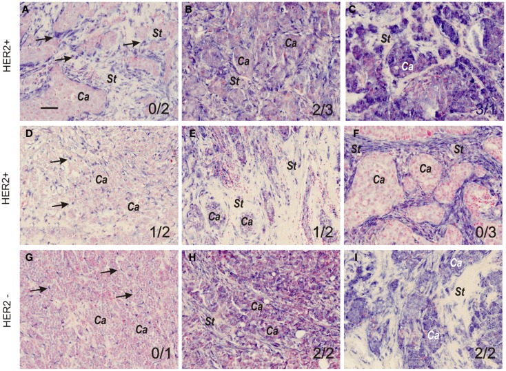
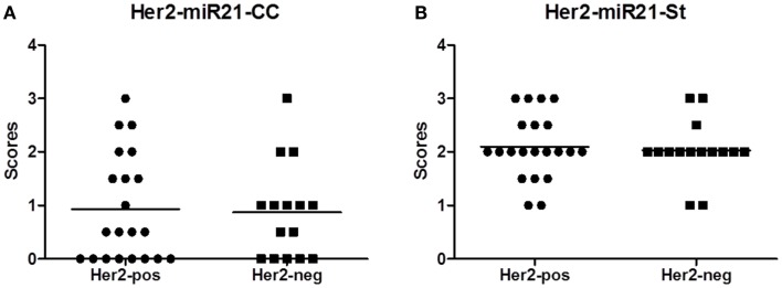
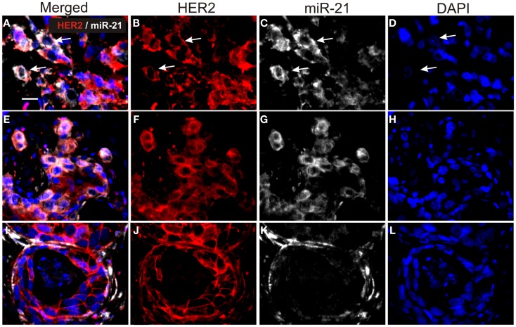
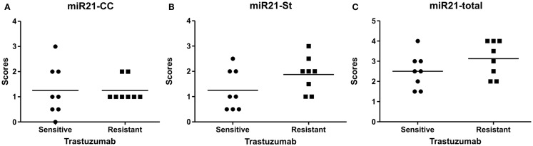
Trastuzumab is established as standard care for patients with HER2-positive breast cancer both in the adjuvant and metastatic setting. However, 50% of the patients do not respond to the trastuzumab therapy, and therefore new predictive biomarkers are highly warranted. MicroRNAs (miRs) constitute a new group of biomarkers and their cellular expression can be determined in tumor samples by in situ hybridization (ISH) analysis. miR-21 is highly prevalent and up-regulated in breast cancer and has been linked to drug resistance in clinical and in vitro settings. To determine expression patterns of miR-21 in high-grade breast cancers, we examined miR-21 expression in 22 HER2-positive tumors and 15 HER2-negative high-grade tumors by ISH. The histological examination indicated that patient samples could be divided into three major expression patterns: miR-21 predominantly in tumor stroma, predominantly in cancer cells, or in both stromal and cancer cells. There was no obvious difference between the HER2-positive and HER2-negative tumors in terms of the miR-21 expression patterns and intensities. To explore the possibility that miR-21 expression levels and/or cellular localization could predict resistance to adjuvant trastuzumab in HER2-positive breast cancer patients, we analyzed additional 16 HER2-positive tumors from patients who were treated with trastuzumab in the adjuvant setting. Eight of the 16 patients showed clinical recurrence and were considered resistant. Examination of the miR-21 expression patterns and intensities revealed no association between the miR-21 scores in the cancer cell population (p = 0.69) or the stromal cells population (p = 0.13) and recurrent disease after adjuvant trastuzumab. Thus, our findings show that elevated miR-21 expression does not predict resistance to adjuvant trastuzumab.
Keywords: HER2; biomarker; breast cancer; miR-21; prediction; response; trastuzumab.
Figures
Similar articles
-
MicroRNA-21 links epithelial-to-mesenchymal transition and inflammatory signals to confer resistance to neoadjuvant trastuzumab and chemotherapy in HER2-positive breast cancer patients.Oncotarget. 2015 Nov 10;6(35):37269-80. doi: 10.18632/oncotarget.5495. Oncotarget. 2015. PMID: 26452030 Free PMC article.
-
Reactive stroma and trastuzumab resistance in HER2-positive early breast cancer.Int J Cancer. 2020 Jul 1;147(1):266-276. doi: 10.1002/ijc.32859. Epub 2020 Jan 22. Int J Cancer. 2020. PMID: 31904863
-
Exosomal miR-1246 and miR-155 as predictive and prognostic biomarkers for trastuzumab-based therapy resistance in HER2-positive breast cancer.Cancer Chemother Pharmacol. 2020 Dec;86(6):761-772. doi: 10.1007/s00280-020-04168-z. Epub 2020 Oct 17. Cancer Chemother Pharmacol. 2020. PMID: 33068176
-
Optimal adjuvant treatment for patients with HER2-positive breast cancer in 2015.Breast. 2015 Nov;24 Suppl 2:S143-8. doi: 10.1016/j.breast.2015.07.034. Epub 2015 Aug 5. Breast. 2015. PMID: 26255196 Review.
-
Overview of resistance to systemic therapy in patients with breast cancer.Adv Exp Med Biol. 2007;608:1-22. doi: 10.1007/978-0-387-74039-3_1. Adv Exp Med Biol. 2007. PMID: 17993229 Review.
Cited by
-
Molecular pathways involved in microRNA-mediated regulation of multidrug resistance.Mol Biol Rep. 2018 Dec;45(6):2913-2923. doi: 10.1007/s11033-018-4358-6. Epub 2018 Sep 7. Mol Biol Rep. 2018. PMID: 30194558 Review.
-
The Role of Non-Coding RNAs in Breast Cancer Drug Resistance.Front Oncol. 2021 Sep 13;11:702082. doi: 10.3389/fonc.2021.702082. eCollection 2021. Front Oncol. 2021. PMID: 34589423 Free PMC article. Review.
-
MicroRNA Biomarkers in IBD-Differential Diagnosis and Prediction of Colitis-Associated Cancer.Int J Mol Sci. 2020 Oct 24;21(21):7893. doi: 10.3390/ijms21217893. Int J Mol Sci. 2020. PMID: 33114313 Free PMC article. Review.
-
Drug-diagnostics co-development in oncology.Front Oncol. 2014 Aug 4;4:208. doi: 10.3389/fonc.2014.00208. eCollection 2014. Front Oncol. 2014. PMID: 25136515 Free PMC article. No abstract available.
-
Functional miRNAs in breast cancer drug resistance.Onco Targets Ther. 2018 Mar 19;11:1529-1541. doi: 10.2147/OTT.S152462. eCollection 2018. Onco Targets Ther. 2018. PMID: 29593419 Free PMC article. Review.
References
LinkOut - more resources
Full Text Sources
Other Literature Sources
Research Materials
Miscellaneous