. 2014:2014:310215.
doi: 10.1155/2014/310215. Epub 2014 Aug 7.

Characterization of glial cell models and in vitro manipulation of the neuregulin1/ErbB system

Affiliations

Characterization of glial cell models and in vitro manipulation of the neuregulin1/ErbB system

Davide Pascal et al. Biomed Res Int. 2014.

Abstract

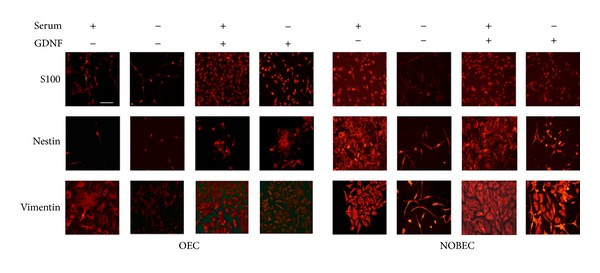
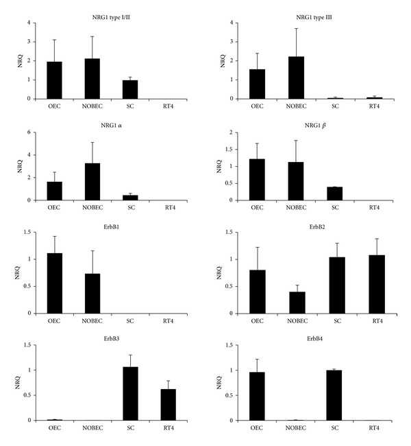
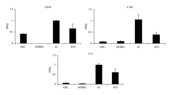
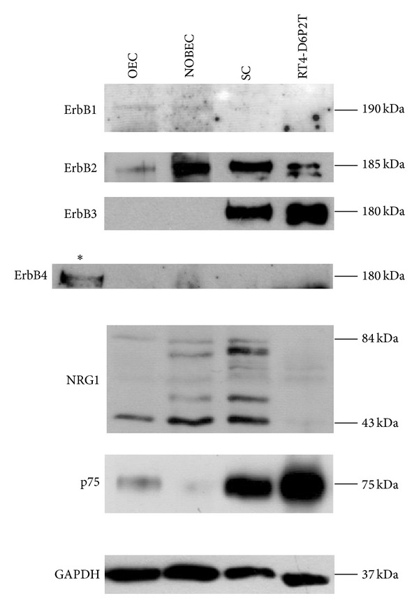
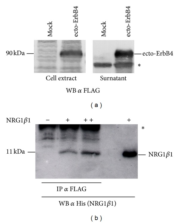
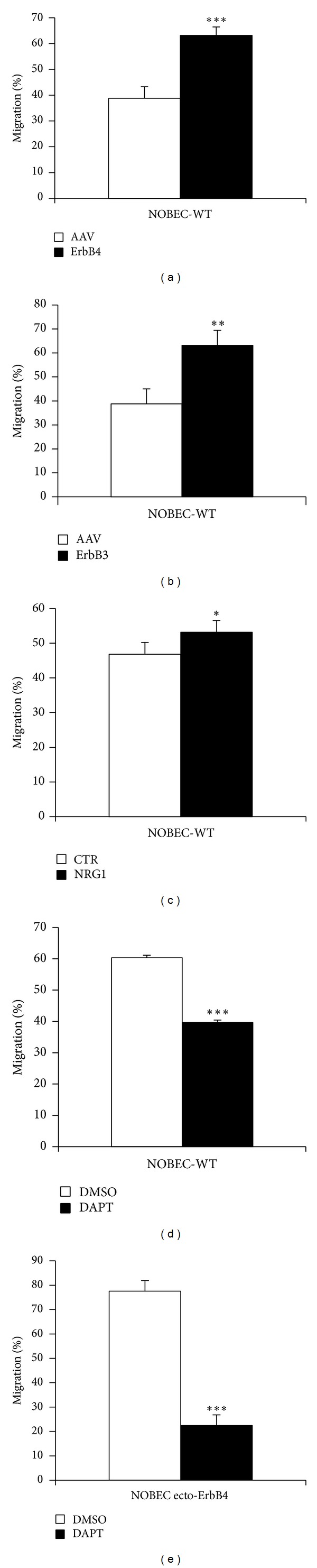
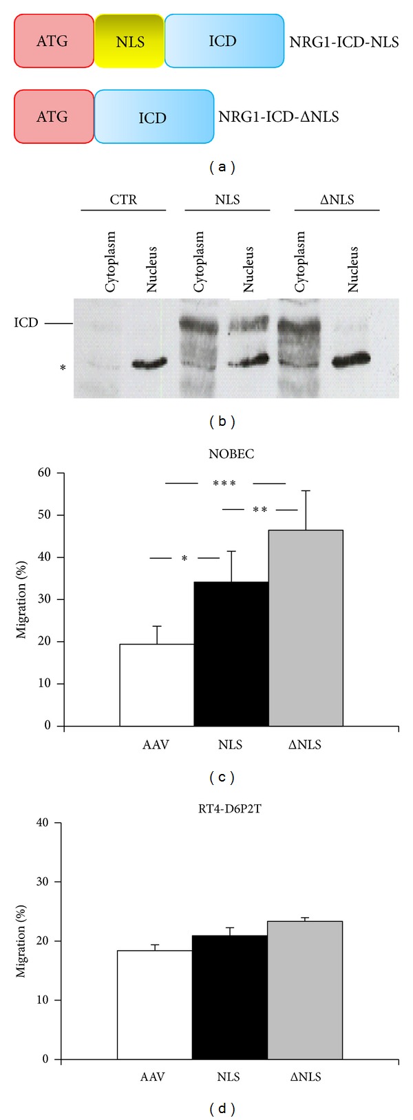
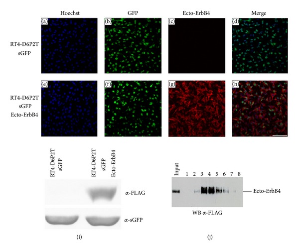
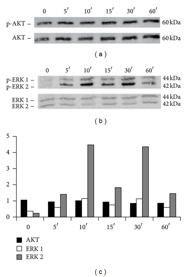
The neuregulin1/ErbB system plays an important role in Schwann cell behavior both in normal and pathological conditions. Upon investigation of the expression of the neuregulin1/ErbB system in vitro, we explored the possibility to manipulate the system in order to increase the migration of Schwann cells, that play a fundamental role in the peripheral nerve regeneration. Comparison of primary cells and stable cell lines shows that both primary olfactory bulb ensheathing cells and a corresponding cell line express ErbB1-ErbB2 and neuregulin1, and that both primary Schwann cells and a corresponding cell line express ErbB2-ErbB3, while only primary Schwann cells express neuregulin1. To interfere with the neuregulin1/ErbB system, the soluble extracellular domain of the neuregulin1 receptor ErbB4 (ecto-ErbB4) was expressed in vitro in the neuregulin1 expressing cell line, and an unexpected increase in cell motility was observed. In vitro experiments suggest that the back signaling mediated by the transmembrane neuregulin1 plays a role in the migratory activity induced by ecto-ErbB4. These results indicate that ecto-ErbB4 could be used in vivo as a tool to manipulate the neuregulin1/ErbB system.


Figures

Figure 1
Figure 1
OEC and NOBEC respond similarly to GDNF stimulation. Figure shows representative fields of OEC and NOBEC immuno-stained with anti-S-100, antinestin, and antivimentin antibodies. Cells were grown for eight days after plating with and without serum and GDNF. Fields were chosen to clearly to show both the morphological aspect and the specific marker expression. Scale bars 50 μm.
Figure 2
Figure 2
Primary cultures and cell lines derived from OEC and SC express different levels of NRG1 isoforms and ErbB receptors. Graphs show normalized relative quantification (NRQ) of the different NRG1 isoforms and ErbB receptors obtained by qRT-PCR. For each gene, the cells with the highest level of expression were chosen as calibrator (NRQ = 1). Data are presented as mean + SEM.
Figure 3
Figure 3
Primary cultures and cell lines derived from OEC and SC express different levels of glial genes. Graphs show normalized relative quantification (NRQ) of GFAP, S100, and p75 obtained by qRT-PCR. For each gene, the cells with the highest level of expression were chosen as calibrator (NRQ = 1). Data are presented as mean + SEM.
Figure 4
Figure 4
Western blot analysis confirms that primary cultures and cell lines derived from OEC and SC express different levels of ErbB receptors and glial proteins. Western blot analysis of proteins extracted from OEC, NOBEC, RT4-D6P2T, and SC were analyzed with antibodies directed to ErbB1, ErbB2, ErbB3, ErbB4, NRG1, p75, and GAPDH. Different NRG1 isoforms are expressed by OEC, NOBEC, and SC. An asterisk indicates a positive control for ErbB4 expression (a cell line stably expressing ErbB4 [18]).
Figure 5
Figure 5
Recombinant ecto-ErbB4-FLAG is expressed and released by cells and is able to interact with soluble NRG1. (a) The correct expression of ecto-ErbB4-FLAG was assayed both in the cell extract and in the conditioned medium (surnatant) of transiently transfected COS7 cells (ecto-ErbB4). Mock samples are COS7 cells transfected with the empty vector. The asterisk indicates an unspecific band. (b) Different amounts of soluble recombinant NRG1β1 tagged with 6 histidine were incubated with conditioned medium of COS7 cells transiently expressing recombinant ecto-ErbB4-FLAG. A FLAG coimmunoprecipitation was performed, followed by Western blot against histidine to recognize NRG1β1. The asterisk indicates the band corresponding to the primary antibody used for coimmunoprecipitation.
Figure 6
Figure 6
NOBEC transiently expressing ecto-ErbB4 and ecto-ErbB3 migrate more than control cells and their migration is inhibited by DAPT treatment. Migration activity of NOBEC transiently transfected with empty vector (AAV) was compared with migration activity of NOBEC expressing ecto-ErbB4 (ErbB4, Panel (a)) or ecto-ErbB3 (ErbB3, Panel (b)). Soluble recombinant NRG1β1 stimulates NOBEC wild type (WT) migration (Panel (c)). NOBEC wild type (Panel (d)) or NOBEC expressing ecto-ErbB4 (Panel (e)) were pretreated for three days with 100 μM DAPT (γ-secretase inhibitor) or DMSO (mock control). Cell migration was assessed by Transwell assays. Values represent the average of three biological replicates performed as technical triplicates. Values of each replicate are expressed in percentage with respect to the total number of cells that migrated in that experiment (∗∗, P ≤ 0.01; ∗∗∗, P ≤ 0.001).
Figure 7
Figure 7
The expression of NRG1 intracellular domain stimulates cell migration. (a) To express the NRG1 intracellular domain (ICD), two constructs were prepared: one containing the nuclear localization sequence (NLS) and one lacking it (ΔNLS). ICD is shown in blue, NLS in yellow, and the inserted ATG in red. (b) Validation of nuclear and cytoplasmic localization of NRG1-ICD fragments. Nuclear and cytoplasmic proteins were extracted from mock (CTR) and NRG1 transfected COS7 cells and subjected to SDS-PAGE and Western blot analysis. Membranes were incubated with anti-NRG1 (sc-348) antibody. Asterisk indicates an unspecific band. (c) NOBEC transiently expressing the NRG1 intracellular domain (ICD), containing (NLS) or lacking (ΔNLS) the nuclear localization sequence, were assayed for migration activity; data show that the cytoplasmic protein confers a migratory activity higher than the migratory activity conferred by the nuclear protein. (d) RT4-D6P2T cells transiently expressing the NRG1 intracellular domain (ICD), containing (NLS) or lacking (ΔNLS) the nuclear localization sequence, were assayed for migration activity. No statistical difference between cells transfected with the empty vector and cells transfected with the two constructs was observed. Values represent the average of three biological replicates performed as technical triplicates. Values of each replicate are expressed in percentage with respect to the total number of cells that migrated in that experiment (∗, P ≤ 0.01; ∗∗, P ≤ 0.01; ∗∗∗, P ≤ 0.001).
Figure 8
Figure 8
Following lentivirus (LV) infection, RT4-D6P2T successfully express GFP and ecto-ErbB4-FLAG. Confluent cells infected with LV-sGFP only (Panels (a)–(d)) or with LV-sGFP and LV-ecto-ErbB4-FLAG (Panels (e)–(h)) were stained with anti-GFP (green) and anti-FLAG (red) antibody. Nuclei were stained with Hoechst (blue). Scale bar 100 μm. Panel (i)-Western blot analysis of RT4-D6P2T infected with LV-sGFP only, or with LV-sGFP and LV-ecto-ErbB4-FLAG. (j) Recombinant ecto-ErbB4-FLAG peptide was purified from RT4-D6P2T conditioned medium using ANTI-FLAG affinity gel and eluted using FLAG peptide. Eluted fractions were analyzed by Western blot to identify the positive fractions to be frozen and used for the following experiments.
Figure 9
Figure 9
Ecto-ErbB4 stimulates ERK phosphorylation in NOBEC. Western blot analysis of NOBEC cells stimulated with recombinant soluble ecto-ErbB4-FLAG for 0, 5, 10, 15, 30, and 60 min. Western blot was analyzed with antibodies anti-p-AKT and AKT (Panel (a)) and anti-p-ERK 1/2 and ERK (Panel (b)). (c) Bands were analyzed by quantifying the intensity of the pixels per mm2 (Image J). The values of the bands corresponding to phosphorylated proteins were normalized to the intensity of the bands corresponding to the total proteins.

References

    1. Britsch S. The neuregulin-I/ErbB signaling system in development and disease. Advances in Anatomy, Embryology, and Cell Biology. 2007;190:1–65. - PubMed
    1. Fricker FR, Bennett DL. The role of neuregulin-1 in the response to nerve injury. Future Neurology. 2011;6(6):809–822. - PMC - PubMed
    1. Gambarotta G, Fregnan F, Gnavi S, Perroteau I. Neuregulin 1 role in Schwann cell regulation and potential applications to promote peripheral nerve regeneration. International Review of Neurobiology. 2013;108:223–256. - PubMed
    1. Taveggia C, Feltri ML, Wrabetz L. Signals to promote myelin formation and repair. Nature Reviews Neurology. 2010;6(5):276–287. - PMC - PubMed
    1. Yarden Y, Sliwkowski MX. Untangling the ErbB signalling network. Nature Reviews Molecular Cell Biology. 2001;2(2):127–137. - PubMed

Publication types