

TomoPy: a framework for the analysis of synchrotron tomographic data
- PMID: 25178011
- PMCID: PMC4181643
- DOI: 10.1107/S1600577514013939
TomoPy: a framework for the analysis of synchrotron tomographic data
Abstract
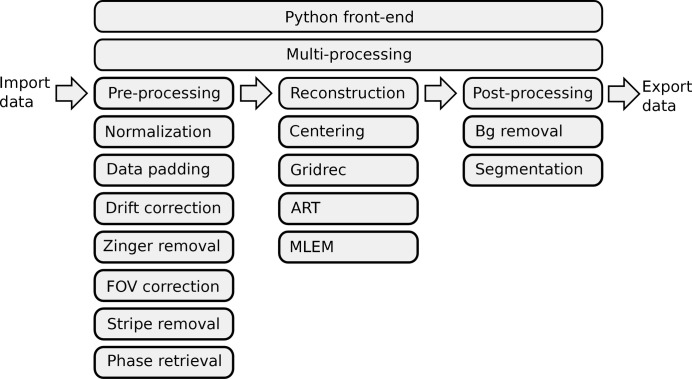
Analysis of tomographic datasets at synchrotron light sources (including X-ray transmission tomography, X-ray fluorescence microscopy and X-ray diffraction tomography) is becoming progressively more challenging due to the increasing data acquisition rates that new technologies in X-ray sources and detectors enable. The next generation of synchrotron facilities that are currently under design or construction throughout the world will provide diffraction-limited X-ray sources and are expected to boost the current data rates by several orders of magnitude, stressing the need for the development and integration of efficient analysis tools. Here an attempt to provide a collaborative framework for the analysis of synchrotron tomographic data that has the potential to unify the effort of different facilities and beamlines performing similar tasks is described in detail. The proposed Python-based framework is open-source, platform- and data-format-independent, has multiprocessing capability and supports procedural programming that many researchers prefer. This collaborative platform could affect all major synchrotron facilities where new effort is now dedicated to developing new tools that can be deployed at the facility for real-time processing, as well as distributed to users for off-site data processing.
Keywords: X-ray imaging; phase retrieval; tomography.
Figures
References
-
- Azevedo, S. G., Schneberk, D., Fitch, J. & Martz, H. (1990). IEEE Trans. Nucl. Sci. 37, 1525–1540.
-
- Banhart, J. (2008). Advanced Tomographic Methods in Materials Research and Engineering. Oxford University Press.
-
- Bronnikov, A. V. (1999). Opt. Commun. 171, 239–244.
-
- Burvall, A., Lundström, U., Takman, P. A. C., Larsson, D. H. & Hertz, H. M. (2011). Opt. Express, 19, 10359–10376. - PubMed
-
- Davis, T. J., Gao, D., Gureyev, T. E., Stevenson, A. W. & Wilkins, S. W. (1999). Nature (London), 373, 595–598.
Publication types
LinkOut - more resources
Full Text Sources
Other Literature Sources
