

Gestational pemphigoid
- PMID: 25178359
- PMCID: PMC4154519
- DOI: 10.1186/s13023-014-0136-2
Gestational pemphigoid
Abstract
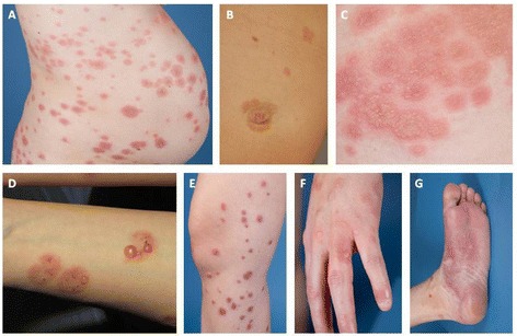
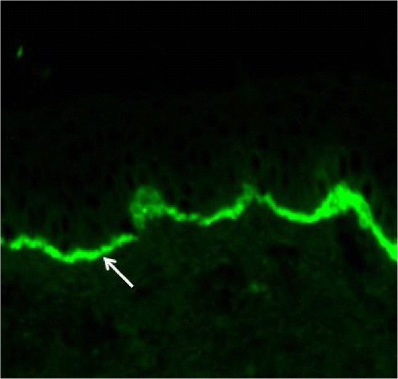
Gestational pemphigoid (pemphigoid gestationis, PG) is a rare autoimmune skin disorder occurring characteristically during pregnancy. Autoantibodies against placental BP180 (also known as BPAG2 or collagen XVII) cause damage to the skin basement membrane, resulting in severe itching and blistering rash over the body and the extremities. The diagnosis of PG is confirmed by immunofluorescence analysis of a skin biopsy, while serum levels of pemphigoid antigen BP180 antibody can be used to assess disease activity. PG with mild symptoms can be treated with topical corticosteroids, while oral corticosteroids are the mainstay in treatment of severe PG. PG usually flares up at the time of delivery, and resolves spontaneously shortly after. However, relapses in subsequent pregnancies are common. As PG has been linked to the risk of prematurity and fetal growth restriction, prenatal monitoring jointly by a dermatologist and an obstetrician is recommended. Mothers should also be informed of the potential risk of re-activation of the disease in subsequent pregnancies and during hormonal contraception.
Figures
References
-
- Schmidt E, Zillikens D. Pemphigoid diseases. Lancet. 2013;381(9863):320–332. - PubMed
-
- Holmes RC, Black MM. The specific dermatoses of pregnancy. J Am Acad Dermatol. 1983;8(3):405–412. - PubMed
-
- Nanda A, Dvorak R, Al-Saeed K, Al-Sabah H, Alsaleh QA. Spectrum of autoimmune bullous diseases in Kuwait. Int J Dermatol. 2004;43(12):876–881. - PubMed
-
- Bernard P, Vaillant L, Labeille B, Bedane C, Arbeille B, Denoeux JP, Lorette G, Bonnetblanc JM, Prost C. Incidence and distribution of subepidermal autoimmune bullous skin diseases in three French regions. Bullous Diseases French Study Group. Arch Dermatol. 1995;131(1):48–52. - PubMed
-
- Bertram F, Brocker E, Zillikens D, Schmidt E. Prospective analysis of the incidence of autoimmune bullous disorders in Lower Franconia, Germany. J Dtsch Dermatol Ges. 2009;7(5):434–439. - PubMed
Publication types
MeSH terms
Substances
LinkOut - more resources
Full Text Sources
Other Literature Sources
Medical
