Review
. 2015 May;16(4):413-34.
doi: 10.1111/mpp.12190. Epub 2014 Dec 11.

The Top 10 oomycete pathogens in molecular plant pathology

Affiliations
Review

The Top 10 oomycete pathogens in molecular plant pathology

Sophien Kamoun et al. Mol Plant Pathol. 2015 May.

Abstract

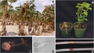
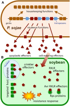
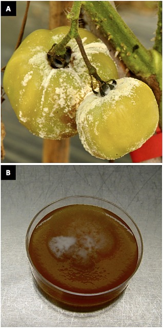
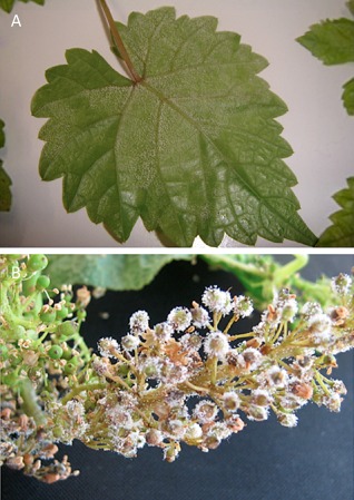
Oomycetes form a deep lineage of eukaryotic organisms that includes a large number of plant pathogens which threaten natural and managed ecosystems. We undertook a survey to query the community for their ranking of plant-pathogenic oomycete species based on scientific and economic importance. In total, we received 263 votes from 62 scientists in 15 countries for a total of 33 species. The Top 10 species and their ranking are: (1) Phytophthora infestans; (2, tied) Hyaloperonospora arabidopsidis; (2, tied) Phytophthora ramorum; (4) Phytophthora sojae; (5) Phytophthora capsici; (6) Plasmopara viticola; (7) Phytophthora cinnamomi; (8, tied) Phytophthora parasitica; (8, tied) Pythium ultimum; and (10) Albugo candida. This article provides an introduction to these 10 taxa and a snapshot of current research. We hope that the list will serve as a benchmark for future trends in oomycete research.

Keywords: diversity; genomics; microbiology; oomycetes plant pathology.


Figures

Figure 1
Figure 1
Potato plants with typical late blight lesions. Infection starts when a spore lands on the leaf and germinates. The germ tube forms an appressorium and an emerging penetration peg pushes into an epidermal cell. Then the inner cell layers are colonized. During the biotrophic phase, hyphae grow in the intercellular space, whereas haustoria enter plant cell cavities and invaginate host cell plasma membrane. Later, Phytophthora infestans switches to necrotrophic growth, resulting in the death of plant cells and the appearance of necrotic lesions on the infected tissues. In this phase, hyphae escape through the stomata and produce numerous asexual spores, named sporangia, that easily detach and disperse by wind or water. A sporangium that finds a new host can either germinate directly and initiate a new cycle or, at lower temperatures, undergo cleavage resulting in a zoosporangium from which six to eight flagellated spores are released. These zoospores can swim for several hours but, once they touch a solid surface, they encyst and germinate to initiate new infections. Under favourable conditions, the pathogen can complete the cycle from infection to sporulation in 4 days. In the field, this cycle is repeated multiple times during one growing season, resulting in billions of spores and a continuous increase in disease pressure. In addition to leaves, stems and tubers are also infected and P. infestans can continue to flourish on the decaying plant material. If not managed properly, infected seed potatoes or waste on refuse piles are often the sources of inoculum for new infections in the spring. An alternative route for surviving the winter is via oospores, sexual spores that can survive in the soil for many years. Phytophthora infestans is heterothallic; isolates are either A1 or A2 mating type, and sex organs only develop when isolates of opposite mating type sense the sex hormone produced by the mate.
Figure 2
Figure 2
One of the many Great Famine memorials around the world. These sculptures on Customs House Quays in Dublin, by artist Rowan Gillespie, stand as if walking towards the emigration ships on the Dublin Quayside (courtesy of Michael Seidl).
Figure 3
Figure 3
Hyaloperonospora arabidopsidis disease symptoms on a 2‐week‐old Arabidopsis seedling. Mature sporangiophores are visible as white structures on the right side of the leaf.
Figure 4
Figure 4
Diagram depicting a compatible interaction between Hyaloperonospora arabidopsidis and Arabidopsis initiated by a sporangiospore landing on the leaf surface. A, appressorium; C, cuticle; Ha, haustorium; Hy, hyphae; LE, lower epidermis; N, nucleus; PM, palisade mesophyll cells; S, sporangiospore; SM, spongy mesophyll cells; Sp, mature sporangiophore; UE, upper epidermis. Note: sporangiophores are not drawn to scale.
Figure 5
Figure 5
Impact of sudden oak death in California. Tanoak mortality evidenced by defoliated or wilted canopies on the Bolinas Ridge at Mt. Tamalpais, Marin County, CA, USA. Photograph courtesy of Janet Klein (Marin Municipal Open Space District).
Figure 6
Figure 6
Inferred pattern of migration of the four clonal lineages of Phytophthora ramorum. Modified from Grünwald et al. (2012).
Figure 7
Figure 7
Silencing machinery in Phytophthora. Phylogenetic placement of dicer‐like (DCL) (A) and argonaute (Ago) (B) proteins in the genus Phytophthora. For more details, see Fahlgren et al. (2013). Species correspond to: Arabidopsis thaliana, Paramecium tetraurelia, Phytophthora infestans, Phytophthora ramorum, Phytophthora sojae, Tetrahymena thermophila and Toxoplasma gondii.
Figure 8
Figure 8
Phytophthora sojae. (A) Diseased soybean plants in the field, infected with P. sojae. Plant height is 20–30 cm. (B) Susceptible (left) and resistant (right) soybean plants inoculated in the stem with P. sojae, 7 days after infection, illustrating R‐gene‐mediated resistance. Pots are 10 cm in diameter. (C) Germinating oospore of P. sojae growing on water agar. Oospore is 35 μm in diameter. (D) Germinating P. sojae cysts growing on water agar. Cysts are 15 μm in diameter. (E) Etiolated soybean hypocotyls inoculated with a 10‐μL water droplet containing 103 zoospores from a virulent (top) and avirulent (bottom) strain of P. sojae, 48 h after infection, illustrating strain‐specific variation in avirulence effectors and the hypersensitive response. Soybean hypocotyls are 5 mm in diameter.
Figure 9
Figure 9
Large diverse families of virulence proteins encoded by the Phytophthora sojae genome. (A) The Phytophthora sojae genome contains clusters of conserved housekeeping genes (brown) that have conserved orders among Phytophthora species, separated by dynamic transposon‐rich regions that contain genes (red) encoding virulence proteins, many of which are secreted. (B) Secreted virulence proteins (effectors) may act in the apoplast, or be transported inside the cell. Cell‐entering effectors may have targets in the nucleus or cytoplasm, and may be detected by resistance proteins (Rps; resistance against P. sojae).
Figure 10
Figure 10
Heavy sporulation and spontaneous morphological variation in the vegetable pathogen Phytophthora capsici. (A) Naturally infected tomato fruit with sporangium production on the surface of the fruit. (B) A single zoospore‐derived field isolate of P. capsici sectoring on V8 agar medium following long‐term storage.
Figure 11
Figure 11
Downy mildew symptoms with well‐evident sporangiophores on the lower side of a grape leaf (A) and a young cluster (B).
Figure 12
Figure 12
Cryo‐scanning electron micrograph of a Phytophthora cinnamomi cyst between two epidermal cells on a root of tobacco. Note the adhesive material that surrounds the cyst that has been expelled by zoospore peripheral vesicles. Photograph courtesy of Adrienne Hardham, Australian National University.
Figure 13
Figure 13
(A) Individual plants of Xanthorrheoa australis (austral grass tree), a highly suscpetible native Australian species, infected by Phytophthora cinnamomi within a dry sclerophyll eucalypt forest at Anglesea, Victoria, Australia. Note the dead and dying plants that have brown, collapsed leaves compared with the healthy green and erect leaves of plants which are yet to be killed. These individual plants range in age from approximately 20 years (the smallest in the centre) to around 70 years (green individual on the right of the image). (B) Advancing disease front caused by invasion by P. cinnamomi in Xanthorrhoea australis‐dominated understorey in eucalyptus open forest at Wilsons Promontory, Victoria, Australia. The disease has moved from the foregound of the picture, where all susceptible vegetation including X. australis has been killed, and its progress can be seen as a line of dead and dying X. australis (brown collapsed plants) at the disease margin. Healthy green plants behind them will soon be killed. Loss of the major understorey components, as in the forground, results in complete structural change and loss of all susceptible species.
Figure 14
Figure 14
Impact of Phytophthora parasitica infection on citrus plants. (A, B) Five‐year‐old citrus plants not infected and infected, respectively, with Phytophthora parasitica. (C, D) Symptoms of P. parasitica on stems: (C) not infected; (D) infected plant displaying gummosis symptoms. (E, F) Leaves and fruits of infected (left) and healthy (right) plants. (G, H) Scanning electron microscope images of citrus fine roots infected with P. parasitica. Yellow arrows show encysted zoospores and germ tube of P. parasitica. Bar represents 20 μm. (A–F) Photographs courtesy of R. J. D. Dalio. (G, H) Photographs courtesy of M. E. Escanferla.
Figure 15
Figure 15
Severe infection of brinjal fruit with Phytophthora parasitica. Typical symptoms are brown, soft, water‐soaked patches which rapidly cover the whole fruit. Brinjal is also known as aubergine or eggplant (Solanum melongena). Photograph courtesy of S. Guha Roy.
Figure 16
Figure 16
Pythium ultimum var. ultimum. (A) Pre‐emergence damping‐off in okra, resulting in the death of most plants at the front of the row. At the back, disease was controlled using metalaxyl. (B) Terminal hyphal swellings; bar, 10 μm. (C) Oospore within oogonium; bar, 10 μm. Images adapted from Kida et al. (2006) with permission.
Figure 17
Figure 17
Disease symptoms of Albugo infections. (A) Disease resulting from the infection of Brassica juncea with an unknown Albugo species. The misshapen inflorescence phenotype is known as a ‘staghead’. (B) An example of immunity suppression by Albugo laibachii: A. thaliana Col‐0 is resistant to Hyaloperonospora arabidopsidis Emoy2 via RPP1 but, when pre‐infected with A. laibachii, can support the growth of both pathogens.
Figure 18
Figure 18
Albugo spp. have compact genomes. Synteny between Albugo laibachii, Pythium ultimum, Hyaloperonospora arabidopsidis and Phytophthora infestans. The region shown is an example of the relatively dense clustering of genes in Albugo species. With increasing genome size, the distance between both genes increases and re‐organizations occur (red, synteny without inversion; blue, inverted regions). Reproduced from Kemen et al. (2011).

References

    1. Adhikari, B.N. , Hamilton, J.P. , Zerillo, M.M. , Tisserat, N. , Levesque, C.A. and Buell, C.R. (2013) Comparative genomics reveals insight into virulence strategies of plant pathogenic oomycetes. PLoS ONE, 8, e75072. - PMC - PubMed
    1. Adhikari, T.B. , Liu, J.Q. , Mathur, S. , Wu, C.R.X. and Rimmer, S.R. (2003) Genetic and molecular analyses in crosses of race 2 and race 7 of Albugo candida . Phytopathology, 93, 959–965. - PubMed
    1. Agrios, G.N. (2005) Plant Pathology. Burlington, Massachusetts: Elsevier Academic Press Publications.
    1. Ah Fong, A.M.V. and Judelson, H.S. (2003) Cell cycle regulator Cdc14 is expressed during sporulation but not hyphal growth in the fungus‐like oomycete Phytophthora infestans . Mol. Microbiol. 50, 487–494. - PubMed
    1. Akinsanmi, O.A. and Drenth, A. (2013) Phosphite and metalaxyl rejuvenate macadamia trees in decline caused by Phytophthora cinnamomi . Crop Protect. 53, 29–36.

Publication types

MeSH terms