. 2015 Feb;35(1):10-5.
doi: 10.1111/neup.12144. Epub 2014 Sep 1.

Simplification of the modified Gallyas method

Affiliations

Simplification of the modified Gallyas method

Nobuo Kuninaka et al. Neuropathology. 2015 Feb.

Abstract

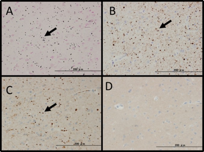
The Gallyas method is a silver impregnation technique that is essential in the field of neuropathology because of its high sensitivity for the detection of argentophilic inclusion bodies in the central nervous system. In Japan, the Gallyas method has improved and is widely used as the "modified Gallyas method". However, this method is not popularly used in general pathology laboratories because of the need for special reagents, several staining processes, and skilled techniques. The objective of the current study was to provide a simplified Gallyas method. We omitted the lanthanum nitrate step from the staining process and verified the adequacy in comparison with the original method as well as immunohistochemistry, using specimens from patients of Alzheimer's disease, argyrophilic grain disease, multiple system atrophy, Pick's disease, and Lewy body disease. The simplified method provided good staining to all the structures in archival tissues, compared with the modified Gallyas method in a significantly shorter staining time. The lanthanum nitrate step can be omitted from the modified Gallyas method, resulting in reduction in the number of reagents required and shortening of the staining time.

Keywords: argyrophilic grains; glial cytoplasmic inclusion; lanthanum nitrate hexahydrate; neurofibrillary tangles; simplification technique.


Figures

Fig. 1
Fig. 1
Suitability of omission of the lanthanum nitrate step (representative cases of corticobasal degeneration in A–C and Alzheimer's disease in D–F). (A):The original Gallyas method. Astrocytic plaque, characteristic of corticobasal degeneration were observed, although the sensitivity was slightly lower (black arrow). (B): The modified Gallyas method. The image shows positive staining with better sensitivity than that of the original. (C): The simplified Gallyas method. The image shows positive staining that was not inferior to that of the modified Gallyas method. Additionally, non-specific background reactivity was not observed. (D): The original method. NFTs and neuropil threads accepted that they are stained with black (black arrow). (E): The modified Gallyas method. The image shows positive staining with better sensitivity than the original, and the absence of non-specific reactions. (F): The simplified Gallyas method. The image shows detection sensitivity that was not inferior to that of the modified Gallyas method, and non-specific background reactivity was not observed. (Scale bar = 200 μm).
Fig. 2
Fig. 2
Influence of time after preparation of the developer solution on the staining efficacy. When the reaction was started 13 min after preparation of the reaction solution, black staining began gradually at 10 min and ended 17 min after the start of staining. When the reaction solution was 40 min old, staining started at 7 min and ended 17 min after the start. When the reaction solution was 60 min old, black staining started at 7 min but ended 15 min after the start. When the reaction solution was 90 min old, staining started at 6 min but ended 13 min after the start. A trend for black staining to start and end earlier in older reaction solutions was observed and occurred in parallel to deterioration of the solution.
Fig. 3
Fig. 3
Comparison between formalin and PFA fixation solutions (representative case: dementia with grains). (A): Formalin fixation. Argyrophilic granules were observed in numbers (black arrows). Non-specific reactions were not observed in peripheral blood vessels, red blood cells, etc. (B): PFA fixation. Although non-specific reactions were observed in peripheral blood vessels and red blood cells, these non-specific reactions did not influence the silver reaction itself (white arrows).
Fig. 4
Fig. 4
Alzheimer's disease. (A): The simplified Gallyas method. Many NFTs including tangles (black arrow) and threads (white arrow) were observed. The specimen was AT8-positive (B), RD3-positive (C), and RD4-positive (D).
Fig. 5
Fig. 5
Dementia with grains. (A): The simplified Gallyas method. Many argyrophilic grains (granules and fusiform structures) (black arrow) were observed. The specimen was AT8-positive (B), RD4-positive (C), and RD3-negative (D).

References

    1. Gallyas F. Silver staining of Alzheimer's neurofibrillary changes by means of physical development. Acta Morphol Acad Sci Hung. 1971;19:1–8. - PubMed
    1. Braak H, Braak E, Ohm T, Bohl J. Silver impregnation of Alzheimer's neurofibrillary changes counterstained for basophilic material and lipofuscin pigment. Stain Technol. 1988;63:197–200. - PubMed
    1. Uchihara T, Kondo H, Kosaka K, Tsukagoshi H. Selective loss of nigral neurons in Alzheimer's disease: a morphometric study. Acta Neuropathol. 1992;83:271–276. Pathology and Clinical Medicine. 1994; 12:163–8. - PubMed
    1. Adachi T, Saito Y, Hatsuta H, et al. Neuropathological asymmetry in argyrophilic grain disease. J Neuropathol Exp Neurol. 2010;69:737–744. - PubMed
    1. Arima K. Brain Banking in Japan: current situation and future prospect. Neuropathology. 2010;30:309.

Publication types

MeSH terms