Review
. 2015 Jul 20;23(3):239-55.
doi: 10.1089/ars.2014.6062. Epub 2014 Oct 9.

Protein Oxidation in Aging: Does It Play a Role in Aging Progression?

Affiliations
Review

Protein Oxidation in Aging: Does It Play a Role in Aging Progression?

Sandra Reeg et al. Antioxid Redox Signal. .

Abstract

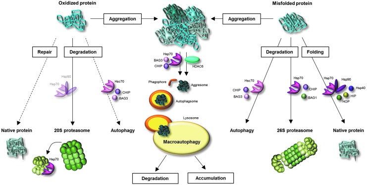
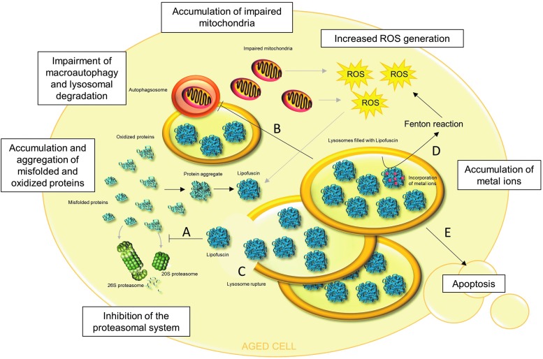
Significance: A constant accumulation of oxidized proteins takes place during aging. Oxidation of proteins leads to a partial unfolding and, therefore, to aggregation. Protein aggregates impair the activity of cellular proteolytic systems (proteasomes, lysosomes), resulting in further accumulation of oxidized proteins. In addition, the accumulation of highly crosslinked protein aggregates leads to further oxidant formation, damage to macromolecules, and, finally, to apoptotic cell death. Furthermore, protein oxidation seems to play a role in the development of various age-related diseases, for example, neurodegenerative diseases.

Recent advances: The highly oxidized lipofuscin accumulates during aging. Lipofuscin formation might cause impaired lysosomal and proteasomal degradation, metal ion accumulation, increased reactive oxygen species formation, and apoptosis.

Critical issues: It is still unclear to which extent protein oxidation is involved in the progression of aging and in the development of some age-related diseases.

Future directions: An extensive knowledge of the effects of protein oxidation on the aging process and its contribution to the development of age-related diseases could enable further strategies to reduce age-related impairments. Strategies aimed at lowering aggregate formation might be a straightforward intervention to reduce age-related malfunctions of organs.


Figures

<b>FIG. 1.</b>
FIG. 1.
Methionine oxidation and repair of methionine sulfoxide (MeSO) via the methionine sulfoxide reductases (MSRs), modified according to Hoshi and Heinemann (71). Oxidation of methionine results in MeSO formation. MeSOs may be repaired by MSRs. Reduction of MeSOs by MSRs leads to the oxidation of the catalytic cysteines in the active sites of the MSRs and to the formation of an intramolecular disulfide. MSRs are recycled due to the thioredoxin/thioredoxin reductase system (Trx/TrxR). Further oxidation of MeSOs leads to the generation of nonrepairable methionine sulfones.
<b>FIG. 2.</b>
FIG. 2.
The proteasomal and the lysosomal degradation system. Although there are several proteasome forms, the attention in this review focuses on 20S proteasome and 26S proteasome. The 20S proteasome consists of four homologous rings, with each divided into seven subunits. The two inner rings are formed by the β-subunits and build the catalytic center of the proteasome; the two outer rings are formed by the α-subunits, which enable recognition and access of substrate proteins to the catalytic center. The 20S proteasome is able to recognize unfolded and, therefore, oxidized proteins due to their increased surface hydrophobicity. Binding of two 19S regulators to the 20S proteasome leads to the formation of the 26S proteasome. Each 19S regulator consists of lid and base and is composed of 19 Rpt- and Rpn-subunits. Polyubiquitination of the substrates, via the ubiqutination system, is necessary for the 26S proteasome to recognize the substrate proteins. In contrast to the 20S proteasome, the 26S proteasome degrades substrate proteins in an ATP-dependent way. ATP hydrolysis is responsible for unfolding of substrate proteins. Lysosomal degradation due to cathepsins is a less selective process than the proteasomal degradation. However, the uptake of substrate proteins via autophagy is a somewhat selective and highly controlled process. While microautophagy describes the direct intake of proteins and organelles into the lysosomal lumen, macroautophagy describes the inclusion first into a phagophore, generating an autophagsome that fuses with the lysosome. Proteins that contain a KFERQ-pentapeptide motive are selectively recognized by Hsc70 and transported to the lysosome. Via lysosomal LAMP-2A receptor and lysosomal Hsc70, proteins are translocated into the lysosomal lumen.
<b>FIG. 3.</b>
FIG. 3.
Fates of oxidized and misfolded proteins and involvement of molecular chaperones. Once a protein is oxidatively modified, several pathways might deal with this oxidized protein. A few oxidative protein modifications may be repaired. However, the large part of oxidized proteins undergoes degradation by the proteasomal system. Since oxidized proteins are partially unfolded, they are a suitable substrates for the 20S proteasome. During oxidative stress, the 26S proteasome dissociates into 20S proteasome and 19S regulators. Hsp70 binds the 19S regulator until re-association of 26S proteasome. The involvement of heat shock proteins (HSPs) in transport of oxidized proteins to the 20S proteasome is assumed, but not proved until now. Moreover, there is some evidence for degradation of oxidized proteins via autophagy. Misfolded (not-covalently modified) proteins are recognized by molecular chaperones, leading to protein refolding or degradation. Folding is accomplished due to Hsp70, Hsp90 and co-chaperones Hsp40, HIP, and HOP. Co-chaperones CHIP and BAG1 direct the Hsp70-bound protein to 26S proteasome, where it is degraded. Misfolded proteins may furthermore undergo autophagy. If oxidized and misfolded proteins are not degraded, repaired, or refolded, they undergo aggregation reactions. Protein aggregates are poor substrates for the proteasome. They generate pericentriolar located aggresomes due to involvement of either histone deacetylase 6 (HDAC6) or Hsp70 and BAG3. The aggresomes may be enclosed by the phagophore, generating the autophagosome, which merges with the lysosome. Degradable proteins are then destroyed by the lysosomal proteases, whereas crosslinked, nondegradable material undergoes further aggregation.
<b>FIG. 4.</b>
FIG. 4.
Human cellular model systems in aging research. Primary cells are often isolated from a young donor. (1) After several subcultures, the young cells reach their replicative limit and are then referred to as senescent postmitotic cells. This process might last several months. The senescent cell stops to divide, but does not immediately die and can be used as a model for aging of nondividing cells. (2) Treatment of young cells with oxidants or stressors for several days/weeks leads to stress-induced premature senescence (SIPS). These cells mirror many features of cells described under (1) and (3). (3) Primary cells isolated from a young donor can be compared with primary cells isolated from an old donor.
<b>FIG. 5.</b>
FIG. 5.
Pathophysiological roles of lipofuscin in aged cells. In many aged cells, the presence of lipofuscin was observed. Lipofuscin is not an inert waste product of cellular metabolism, but exerts multiple effects. (A) Lipofuscin inhibits the 20S and 26S proteasome, resulting in decreased degradation and further accumulation of oxidized and misfolded proteins. This leads to a facilitation of protein aggregate formation and further lipofuscin formation. (B) It is hypothesized that accumulation of lipofuscin in lysosomes may impair autophagy and lysosomal degradation, which may result in decreased degradation of various proteins, protein aggregates, and organelles. Thus, nonfunctional, impaired mitochondria may not be degraded and accumulate, generating increased amounts of reactive oxygen species (ROS). This leads to further protein oxidation, protein aggregation, and lipofuscin formation. (C) Lysosomal membranes are very susceptible to oxidative damage, which may lead to lysosome rupture. This releases lipofuscin into the cytosol, resulting in the consequences described under (A), (D), and (E). (D) Lipofuscin has the ability to incorporate metal ions, which generate highly reactive ROS via the Fenton reaction. (E) An overall consequence of lipofuscin formation and accumulation is apoptotic cell death.
<b>FIG. 6.</b>
FIG. 6.
Role of oxidized low-density lipoprotein (oxLDL) in development of atherosclerosis. Due to an endothelial dysfunction, (modified) low-density lipoprotein (LDL) immigrates from arterial lumen into the arterial wall. In the arterial wall, ROS are generated due to a number of enzymes, including nitric oxide synthase, myeloperoxidase, and NADPH oxidase, and via metal ion-catalyzed Fenton reaction, what results in LDL oxidation (oxLDL). Furthermore, it is still a matter of debate whether circulating oxLDL may also immigrate into the arterial wall. OxLDL stimulates endothelial cells and smooth muscle cells to secrete monocyte chemotactic protein-1 (MCP-1). MCP-1 stimulates arterial monocytes and lymphocytes to immigrate into the arterial wall, where monocytes are converted to macrophages. These macrophages have specific scavenger receptors for oxLDL. Phagocytosis of oxLDL contributes to foam cell formation, which results in plaque formation and thrombosis.

References

    1. Abeliovich A, Schmitz Y, Farinas I, Choi-Lundberg D, Ho WH, Castillo PE, Shinsky N, Verdugo JM, Armanini M, Ryan A, Hynes M, Phillips H, Sulzer D, and Rosenthal A. Mice lacking alpha-synuclein display functional deficits in the nigrostriatal dopamine system. Neuron 25: 239–252, 2000 - PubMed
    1. Aeschbach R, Amado R, and Neukom H. Formation of dityrosine cross-links in proteins by oxidation of tyrosine residues. Biochim Biophys Acta 439: 292–301, 1976 - PubMed
    1. Aksenov MY, Aksenova MV, Butterfield DA, Geddes JW, and Markesbery WR. Protein oxidation in the brain in Alzheimer's disease. Neuroscience 103: 373–383, 2001 - PubMed
    1. Alberti S, Demand J, Esser C, Emmerich N, Schild H, and Hohfeld J. Ubiquitylation of BAG-1 suggests a novel regulatory mechanism during the sorting of chaperone substrates to the proteasome. J Biol Chem 277: 45920–45927, 2002 - PubMed
    1. Antunes F, Cadenas E, and Brunk UT. Apoptosis induced by exposure to a low steady-state concentration of H2O2 is a consequence of lysosomal rupture. Biochem J 356: 549–555, 2001 - PMC - PubMed