

How to interpret postoperative X-rays after total knee arthroplasty
- PMID: 25179351
- PMCID: PMC6583264
- DOI: 10.1111/os.12123
How to interpret postoperative X-rays after total knee arthroplasty
Abstract
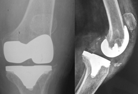
Today, total knee arthroplasty (TKA) is one the most commonly performed surgeries worldwide. The purpose of this article is to review the appearance of normal post-TKA roentgenographs and describe the correct sequence for their interpretation. It is unwise to depend solely on patients' symptoms when diagnosing TKA complications because serial radiographs can foresee failures well before they manifest clinically. Ideal post-TKA radiographs comprise whole lower extremity anteroposterior and lateral views taken under weight bearing conditions along with a skyline view of the patellofemoral joint. Among other things, weight bearing exposes the true alignment, ligamentous laxity and polyethylene wear. On the basis of follow-up of our TKA cases, we have drawn up a protocol for assessing postoperative X-ray films after TKAs. Following the proposed sequence, surgeon can easily decide how to proceed with follow-up and foresee complications. Careful interpretation of postoperative radiographs after TKA is essential to careful monitoring of patients and implant survival.
Keywords: Arthroplasty; Interpretation; Knee; Roentgenography.
© 2014 Chinese Orthopaedic Association and Wiley Publishing Asia Pty Ltd.
Figures


References
-
- Chiu KY, Cheng HC, Yau WP, Tang WM, Ho HS. Reading radiographs after total knee arthroplasty. J Orthop Surg (Hong Kong), 2010, 14: 25–39.
-
- Scuderi GR, Pagnano MW. Review article: the rationale for posterior cruciate substituting total knee arthroplasty. J Orthop Surg (Hong Kong), 2001, 9: 81–88. - PubMed
-
- Barnes CL, Sledge CB. Total knee arthroplasty with posterior cruciate ligament retention designs In: Insall JN, Windsor RE, Scott WN, et al, eds. Surgery of the Knee, 2nd edn New York: Churchill, Livingstone, 1993; 815–827.
-
- Allen AM, Ward WG, Pope TL Jr. Imaging of the total knee arthroplasty. Radiol Clin North Am, 1995, 33: 289–303. - PubMed
-
- Berquist TH. Imaging of joint replacement procedures. Radiol Clin North Am, 2006, 44: 419–437. - PubMed
Publication types
MeSH terms
LinkOut - more resources
Full Text Sources
Other Literature Sources
Medical
