Review
. 2014 Oct 15;127(Pt 20):4343-50.
doi: 10.1242/jcs.154633. Epub 2014 Sep 1.

Defective channels lead to an impaired skin barrier

Affiliations
Review

Defective channels lead to an impaired skin barrier

Diana C Blaydon et al. J Cell Sci. .

Abstract

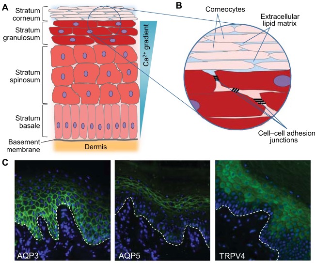
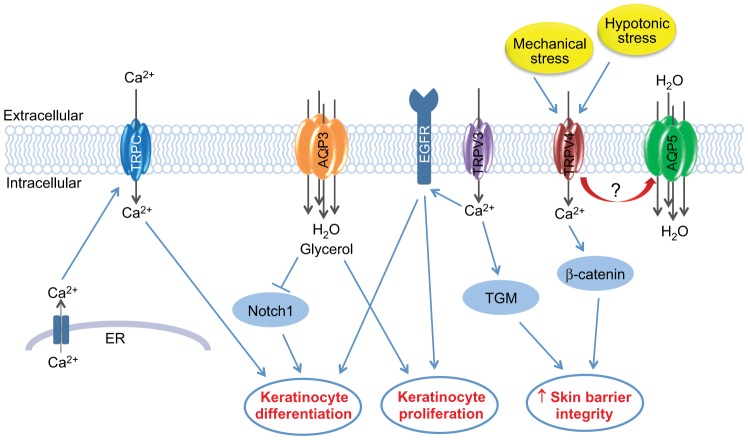
Channels are integral membrane proteins that form a pore, allowing the passive movement of ions or molecules across a membrane (along a gradient), either between compartments within a cell, between intracellular and extracellular environments or between adjacent cells. The ability of cells to communicate with one another and with their environment is a crucial part of the normal physiology of a tissue that allows it to carry out its function. Cell communication is particularly important during keratinocyte differentiation and formation of the skin barrier. Keratinocytes in the skin epidermis undergo a programme of apoptosis-driven terminal differentiation, whereby proliferating keratinocytes in the basal (deepest) layer of the epidermis stop proliferating, exit the basal layer and move up through the spinous and granular layers of the epidermis to form the stratum corneum, the external barrier. Genes encoding different families of channel proteins have been found to harbour mutations linked to a variety of rare inherited monogenic skin diseases. In this Commentary, we discuss how human genetic findings in aquaporin (AQP) and transient receptor potential (TRP) channels reveal different mechanisms by which these channel proteins function to ensure the proper formation and maintenance of the skin barrier.

Keywords: Aquaporin; Gap junction; TRP channel.


Figures

Fig. 1.
Fig. 1.
Structure of the epidermis and epidermal barrier and localisation of AQP3, AQP5 and TRPV4 in normal palmar skin. (A) Illustration of the different layers of keratinocytes found in the epidermis. Keratinocytes in the stratum basale (the deepest layer of the epidermis) are attached to the basement membrane and undergo proliferation. Upon leaving the stratum basale, keratinocytes stop proliferating and enter a programme of terminal differentiation. These cells move up through the stratum spinosum and stratum granulosum layers of the epidermis, undergoing a conformational change as they do so. In the outer layer of the epidermis, the stratum corneum, the keratinocytes are flattened anucleated dead cells known as corneocytes, which are continuously lost from the surface of the skin and replenished by cells from the lower layers of the epidermis. Ca2+ is essential for the regulation of keratinocyte differentiation, and a Ca2+ gradient is seen in the epidermis. (B) The epidermal barrier is formed from a number of components. The stratum corneum forms the main physical barrier of the skin and consists of corneocytes, with a toughened cornified cell envelope, embedded in an extracellular lipid matrix, which forms the main permeability barrier. Cell–cell adhesion junctions, including desmosomal junctions, tight junctions and adherens junctions, between the living nucleated keratinocytes of the lower granular and spinous layers also provide strength to the skin barrier and restrict paracellular permeability. (C) Localisation of some key aquaporin and TRP channel proteins in normal palmar skin is shown using immunofluorescent staining. AQP3 (green, left panel) is expressed in the basal and spinous layers of the epidermis, but AQP3 expression is lost in the upper granular layer, consistent with a role for AQP3 in keratinocyte proliferation and negative regulation of differentiation. By contrast, AQP5 (green, middle panel) shows a strong plasma membrane localisation in keratinocytes of the granular layer, indicating a role for AQP5 in the formation or maintenance of the epidermal barrier, although further work is required to establish the function of AQP5 in the epidermis. Similarly, TRPV4 (green, right panel) is also expressed in the upper layers of the epidermis, where it is has been shown to play a role in the formation of the intercellular junctions in the epidermal barrier. An association between TRPV4 and AQP5 has been demonstrated in other cell types, and they might have a similar relationship in keratinocytes, although further studies are required to establish this. Nuclei are shown in blue (DAPI), and the white dashed line indicates the location of the basement membrane.
Fig. 2.
Fig. 2.
Illustration of the role of key aquaporin and TRP channel proteins in formation of the epidermal barrier. TRP channels form tetrameric cation-permeable pores, which allow the movement of a variety of cations, including Ca2+, which is crucial for the regulation of keratinocyte differentiation. A number of TRPC channels (blue) are expressed in keratinocytes, and there is evidence supporting a key role for these channels in the promotion of Ca2+-induced keratinocyte differentiation. TRPC channels are believed to be activated (thus allowing the influx of extracellular Ca2+) upon detection of Ca2+ release from internal stores, such as the ER. TRPV3 (purple) appears to play a central role in keratinocyte biology, with a key role in establishing the balance between keratinocyte proliferation and differentiation, which is believed to be mediated through EGFR activation. TRPV3 might also have a more direct role in the formation of the epidermal barrier through regulation of the activity of transglutaminases (TGM), the enzymes required for the cross-linking of protein components of the cornified envelope. Furthermore, TRPV4 (red), which is activated in response to mechanical and hypotonic stress, has been shown to bind to components of the intercellular junctions, such as β-catenin, leading to increased skin barrier integrity. AQP3 (orange), an aquaglyceroporin that is abundantly expressed in keratinocytes, also allows the movement of small molecules, such as glycerol, in addition to water, into keratinocytes. AQP3 appears to play a central role in keratinocyte proliferation, and although the role for AQP3 in keratinocyte differentiation is less clear, there is evidence that it might be involved in the negative regulation of differentiation through inhibition of Notch1, a known mediator of keratinocyte differentiation. AQP5 is a water-selective aquaporin, the role of which in the formation of the epidermal barrier is currently unclear. However, AQP5 (green) might have a role in the regulation of cell–cell adhesion, either through microtubule stabilisation or through an association with TRPV4, as has been shown in other cell types, but further work is required to determine this.

References

    1. Ahmad S., Diez J. A., George C. H., Evans W. H. (1999). Synthesis and assembly of connexins in vitro into homomeric and heteromeric functional gap junction hemichannels. Biochem. J. 339, 247–253 10.1042/0264-6021:3390247 - DOI - PMC - PubMed
    1. Akazawa Y., Yuki T., Yoshida H., Sugiyama Y., Inoue S. (2013). Activation of TRPV4 strengthens the tight-junction barrier in human epidermal keratinocytes. Skin Pharmacol. Physiol. 26, 15–21 10.1159/000343173 - DOI - PubMed
    1. Asakawa M., Yoshioka T., Matsutani T., Hikita I., Suzuki M., Oshima I., Tsukahara K., Arimura A., Horikawa T., Hirasawa T. et al. (2006). Association of a mutation in TRPV3 with defective hair growth in rodents. J. Invest. Dermatol. 126, 2664–2672 10.1038/sj.jid.5700468 - DOI - PubMed
    1. Bakirtzis G., Choudhry R., Aasen T., Shore L., Brown K., Bryson S., Forrow S., Tetley L., Finbow M., Greenhalgh D. et al. (2003). Targeted epidermal expression of mutant Connexin 26(D66H) mimics true Vohwinkel syndrome and provides a model for the pathogenesis of dominant connexin disorders. Hum. Mol. Genet. 12, 1737–1744 10.1093/hmg/ddg183 - DOI - PubMed
    1. Baroja-Mazo A., Barberà-Cremades M., Pelegrín P. (2013). The participation of plasma membrane hemichannels to purinergic signaling. Biochim. Biophys. Acta 1828, 79–93 10.1016/j.bbamem.2012.01.002 - DOI - PubMed

Publication types

Substances