Review
. 2014 Sep 1;206(5):579-88.
doi: 10.1083/jcb.201404124.

Assemblages: functional units formed by cellular phase separation

Affiliations
Review

Assemblages: functional units formed by cellular phase separation

Jeffrey A Toretsky et al. J Cell Biol. .

Abstract

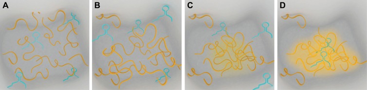
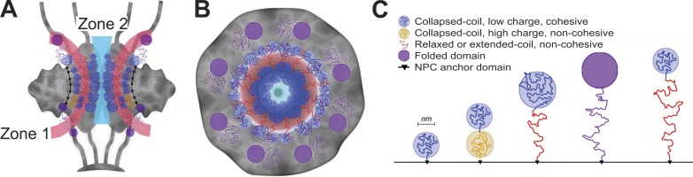
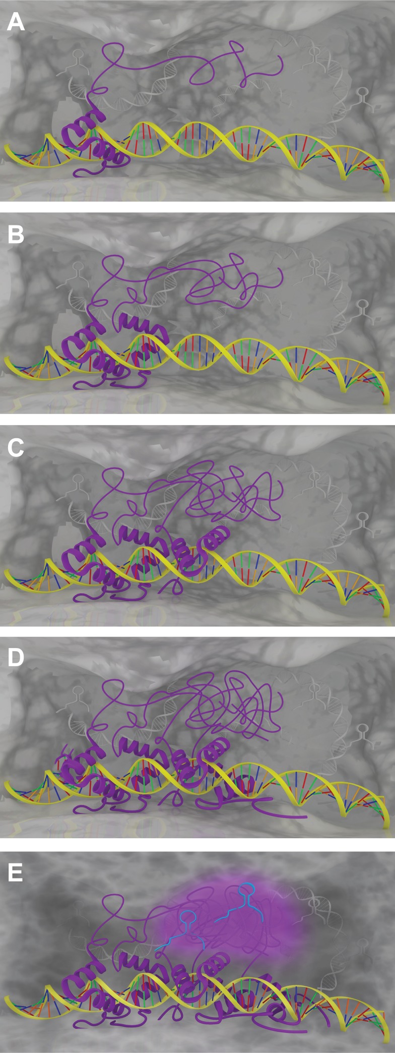
The partitioning of intracellular space beyond membrane-bound organelles can be achieved with collections of proteins that are multivalent or contain low-complexity, intrinsically disordered regions. These proteins can undergo a physical phase change to form functional granules or other entities within the cytoplasm or nucleoplasm that collectively we term "assemblage." Intrinsically disordered proteins (IDPs) play an important role in forming a subset of cellular assemblages by promoting phase separation. Recent work points to an involvement of assemblages in disease states, indicating that intrinsic disorder and phase transitions should be considered in the development of therapeutics.


Figures

Figure 1.
Figure 1.
Assemblage formation leads to emergent properties of protein and RNA binding. This series of panels (A–D) demonstrates that an increase in the local concentration of protein (yellow ribbons) in regions of a cell can result in a phase transition (yellow haze) to form an assemblage once a critical concentration has been reached. A phase separated assemblage can be formed through weak homotypic or heterotypic interactions and allows exchange of constituent molecules with the surrounding solution. This phase-separated material allows for the capture and interaction of other protein or RNA species (cyan molecule). (D) The final assemblage formation shows the sequestration of two RNA molecules.
Figure 2.
Figure 2.
Phase transition caused by IDPs regulates transport in nuclear pore channels. (A) Sagittal plane of a nuclear pore that is populated with IDPs. FG proteins, and variants described in the text, contribute to the “filling” of space between structural protein components (gray). The architecture of the NPC creates zones of transport, indicated here as light red and light blue. (B) Transverse view of nuclear pore complex components (Nups) showing the localization of FG proteins and the transport zones. (C) Legend demonstrating the types of proteins found in the nuclear pore complex and their biophysical characteristics. This figure is based upon Saccharomyces cerevisiae FG Nups and is adapted from Yamada et al. (2010).
Figure 3.
Figure 3.
DNA acts as a scaffold for EWS-FLI1 binding to GGAA repeats, leading to a putative phase transition based upon high concentration of EWS domains. A series of panels (A–D) shows sequential binding of EWS-FLI1 (purple with helical region) to GGAA (red/green) repeats in the DNA. The high concentration of EWS domains that would occur as a result of multiple EWS-FLI1 proteins binding in a DNA microsatellite could lead to a phase transition based upon the intrinsically disordered low-complexity repeats. (E) The increased local concentration of these EWS domain subunits have emergent properties, at a critical concentration depicted here as five proteins, because of a phase transition leading to the sequestration of RNA (cyan). The assemblage and its interaction with RNA could be part of the transcriptional or posttranscriptional machinery. The capture of RNA could tether this dynamic phase separated assemblage to the nascent pre-mRNA or to the posttranscriptional splicing complex.

Similar articles

Cited by

References

    1. Adams, R.L., and Wente S.R.. 2013. Uncovering nuclear pore complexity with innovation. Cell. 152:1218–1221 10.1016/j.cell.2013.02.042 - DOI - PMC - PubMed
    1. Ader, C., Frey S., Maas W., Schmidt H.B., Görlich D., and Baldus M.. 2010. Amyloid-like interactions within nucleoporin FG hydrogels. Proc. Natl. Acad. Sci. USA. 107:6281–6285 10.1073/pnas.0910163107 - DOI - PMC - PubMed
    1. Aggarwal, S., Snaidero N., Pähler G., Frey S., Sánchez P., Zweckstetter M., Janshoff A., Schneider A., Weil M.T., Schaap I.A., et al. . 2013. Myelin membrane assembly is driven by a phase transition of myelin basic proteins into a cohesive protein meshwork. PLoS Biol. 11:e1001577 10.1371/journal.pbio.1001577 - DOI - PMC - PubMed
    1. André, J., and Rouiller C.. 1957. The ultrastructure of the vitelline body in the oocyte of the spider Tegenaria parietina. J. Biophys. Biochem. Cytol. 3:977–984 10.1083/jcb.3.6.977 - DOI - PMC - PubMed
    1. Ano Bom, A.P., Rangel L.P., Costa D.C., de Oliveira G.A., Sanches D., Braga C.A., Gava L.M., Ramos C.H., Cepeda A.O., Stumbo A.C., et al. . 2012. Mutant p53 aggregates into prion-like amyloid oligomers and fibrils: implications for cancer. J. Biol. Chem. 287:28152–28162 10.1074/jbc.M112.340638 - DOI - PMC - PubMed

Publication types

Substances

LinkOut - more resources