. 2015 Jan;244(1):86-97.
doi: 10.1002/dvdy.24185. Epub 2014 Sep 16.

Selective neuronal lineages derived from Dll4-expressing progenitors/precursors in the retina and spinal cord

Affiliations

Selective neuronal lineages derived from Dll4-expressing progenitors/precursors in the retina and spinal cord

Min Zou et al. Dev Dyn. 2015 Jan.

Abstract

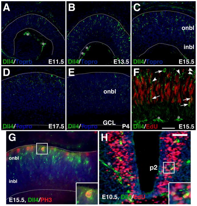
Background: During retinal and spinal cord neurogenesis, Notch signaling plays crucial roles in regulating proliferation and differentiation of progenitor cells. One of the Notch ligands, Delta-like 4 (Dll4), has been shown to be expressed in subsets of retinal and spinal cord progenitors/precursors and involved in neuronal subtype specification. However, it remains to be determined whether Dll4 expression has any progenitor/precursor-specificity contributing to its functional specificity during neural development.

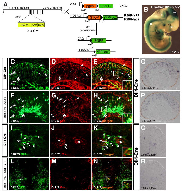
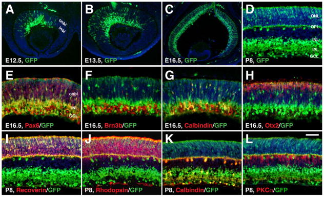
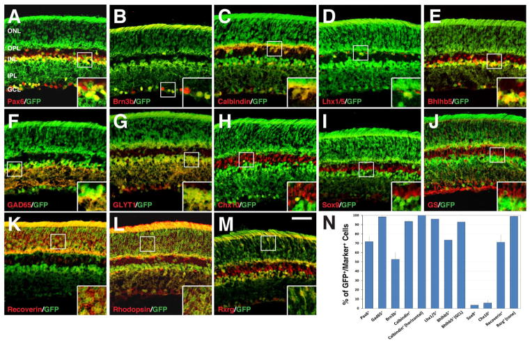
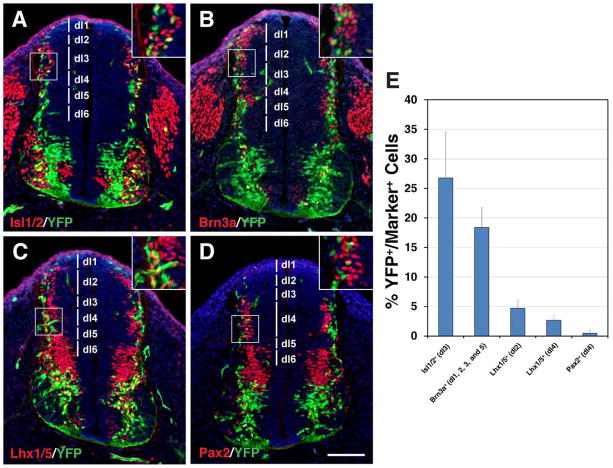
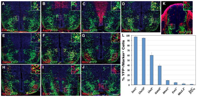
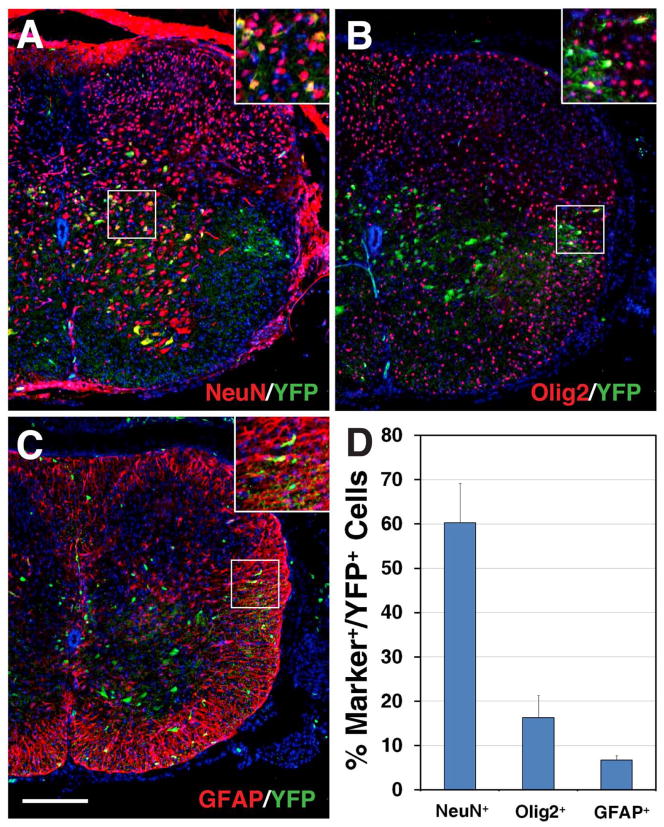
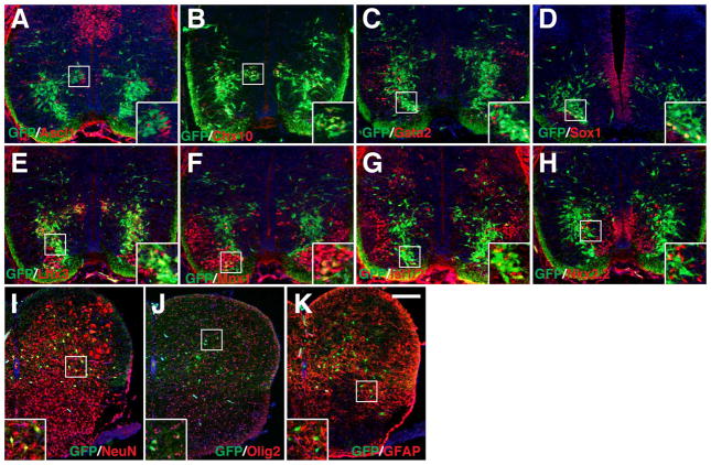
Results: We generated a Dll4-Cre BAC transgenic mouse line that drives Cre recombinase expression mimicking that of the endogenous Dll4 in the developing retina and spinal cord. By fate-mapping analysis, we found that Dll4-expressing progenitors/precursors give rise to essentially all cone, amacrine and horizontal cells, a large portion of rod and ganglion cells, but only few bipolar and Müller cells. In the spinal cord, Dll4-expressing progenitors/precursors generate almost all V2a and V2c cells while producing only a fraction of the cells for other interneuron and motor neuron subtypes along the dorsoventral axis.

Conclusions: Our data suggest that selective expression of Dll4 in progenitors/precursors contributes to its functional specificity in neuronal specification and that the Dll4-Cre line is a valuable tool for gene manipulation to study Notch signaling.

Keywords: Dll4; Notch signaling; V2 interneuron; neuronal lineage; retina; spinal cord.


Figures

Fig. 1
Fig. 1
Expression of Dll4 protein in the developing retina and spinal cord. A–E: Retinal sections from the indicated stages were immunostained with an anti-Dll4 antibody (green), with nuclei counterstained with Topro (blue). The expression of Dll4 starts at E11.5 in the central retina, peaks at E13.5, and remains in the outer neuroblastic layer from E15.5 to P4. F: A retinal section from an E15.5 embryo pulse-labeled with EdU was both chemically stained for EdU (red) and immunostained with an anti-Dll4 antibody (green). Arrows point to representative colocalized cells while arrowheads point to representative cells labeled only for Dll4. G: An E15.5 retinal section was co-immunostained by antibodies against Dll4 and phosphorylated histone H3 (PH3). Note that Dll4 colocalizes with PH3 in scattered cells located at the outer edge of the onbl. H: An EdU pulse-labeled E10.5 spinal cord section was immunostained by Dll4 antibody and chemically stained for EdU. Note that Dll4 colocalizes with EdU in scattered cells around the p2 domain of ventral spinal cord. The asterisks indicate Dll4-immunoreactive blood vessels. Insets show corresponding outlined regions with representative colocalized cells at a higher magnification. Dashed lines outline the thickness of the retina. Abbreviations: GCL, ganglion cell layer; inbl, inner neuroblastic layer; onbl, outer neuroblastic layer. Scale bar in F = 75 μm (A, B), 47.6 μm (D, E), 39.7 μm (C), and 26.5 μm (F); Scale bar in H = 33.3 μm (G) and 50 μm (H).
Fig. 2
Fig. 2
Generation of Dll4-Cre transgenic mice that express Cre recombinase in progenitors/precursors of the retina and spinal cord. A: The Cre-loxP system for conditional activation of reporter YFP/lacZ or GFP expression using Dll4-Cre and R26R-YFP/lacZ or Z/EG mice. To generate the Dll4-Cre BAC, a BAC containing the Dll4 locus was modified by recombineering to insert the Cre coding region (Cre-pA) at the Dll4 translation initiation site (ATG), followed by the E. coli ampicillin resistance gene (Amp) and the R6Kγ origin for DNA replication. The exons are indicated by vertical black and gray bars and the estimated lengths of 5′-flanking and 3′ flanking sequences are also indicated. B: β-gal activity was visualized by X-gal staining in an E12.5 Dll4-Cre; R26R-lacZ wholemount embryo. C–E: A retinal section from an E13.5 Dll4-Cre embryo was immunolabeled with antibodies against Dll4 and Cre. F–H: A retinal section from E12.5 Dll4-Cre; Z/EG embryo was immunostained with antibodies against GFP and Cre. I–K: A spinal cord section from an E10.75 Dll4-Cre embryo was immunolabeled with antibodies against Dll4 and Cre. L–N: A spinal cord section from E12.5 Dll4-Cre; R26R-YFP embryo was immunostained with antibodies against YFP and Cre. Arrows point to representative colocalized cells, arrowheads point to representative non-colocalized cells, and the asterisks in (I, K) indicate Dll4-immunoreactive blood vessels. Insets show corresponding outlined regions at a higher magnification. O–R: RNA in situ hybridization was performed for detecting Dll4 (O, Q) and Cre (P, R) expression on adjacent sections. Abbreviations: a, aorta; e, eye; inbl, inner neuroblastic layer; m, mesencephalon; onbl, outer neuroblastic layer; r, rhombencephalon; s, spinal cord; V2, V2 domain. Scale bar=125 μm (Q, R), 100 μm (LP), and 50 μm (CK).
Fig. 3
Fig. 3
Selective retinal cell types derived from Dll4-expressing progenitors/precursors from E12.5 to P8. A–L: Retinal sections from Dll4-Cre; Z/EG mice at the indicated stages were immunostained with the indicated antibodies. Sections were counterstained with DAPI. Note the colocalization between GFP and Pax6, Brn3b, calbindin, Otx2, recoverin, or rhodopsin, but the absence of colocalization between GFP and PKCα. Abbreviations: GCL, ganglion cell layer; inbl, inner neuroblastic layer; INL, inner nuclear layer; IPL, inner plexiform layer; onbl, outer neuroblastic layer; ONL, outer nuclear layer; OPL, outer plexiform layer. Scale bar= 200 μm (C), 100 μm (A, B), and 50 μm (DL).
Fig. 4
Fig. 4
Retinal cell types derived from Dll4-expressing progenitors/precursors at P21. A–M: P21 retinal sections from Dll4-Cre; Z/EG mice were immunostained with the indicated antibodies. Insets show corresponding outlined regions at a higher magnification. Note the colocalization between GFP and Pax6, Brn3b, calbindin, Lhx1/5, Bhlhb5, GAD65, GLYT1, recoverin, rhodopsin, or Rxrg, but almost absence of colocalization between GFP and Chx10, Sox9, or GS (glutamine synthetase). N: Quantification of marker-immunoreactive cells that are positive for GFP in the Dll4-Cre; Z/EG retina. Each histogram represents the mean±SD for 3 retinas. Note that both total number of immunoreactive cells and immunoreactive cells within layers indicated in the parentheses were counted for calbindin, Bhlhb5 and Rxrg. Abbreviations: GCL, ganglion cell layer; INL, inner nuclear layer; IPL, inner plexiform layer; ONL, outer nuclear layer; OPL, outer plexiform layer. Scale bar = 47.6 μm (AM).
Fig. 5
Fig. 5
Dll4-expressing progenitors/precursors give rise to selective dorsal interneuron lineages in the spinal cord. A–D: Spinal cord sections from E10.75 Dll4-Cre; R26R-YFP embryos were stained by double-immunofluorescence using the indicated antibodies, as well as weakly counterstained with nuclear DAPI. Insets show corresponding outlined regions with representative colocalized cells at a higher magnification. YFP colocalizes with Isl1/2, Brn3a and Lhx1/5 but rarely with Pax2 in distinct domains (indicated by vertical lines) in the dorsal spinal cord. E: Percentages of dorsal interneuron marker-positive cells that are immunoreactive for YFP at E10.75. Each histogram represents the mean±SD for 3 different spinal cords. Note that each marker was counted within their distinct domains as indicated in the parentheses. Scale bar = 100 μm.
Fig. 6
Fig. 6
Selective ventral spinal cord cell types derived from Dll4-expressing progenitors/precursors. A–K: Spinal cord sections from E12.5 (AJ) or E10.75 (K) Dll4-Cre; R26R-YFP embryos were stained by double-immunofluorescence using the indicated antibodies as well as weakly counterstained with nuclear DAPI. Insets show corresponding outlined regions with representative colocalized cells at a higher magnification. Note that YFP colocalizes with Cre in many cells in the p2/V2 domain. YFP colocalizes also with Lhx3, Chx10, Gata2, Sox1, Mnx1, and Isl1/2 but rarely with Ascl1, Evx1, Nkx2.2 or En1 in distinct domains of the ventral spinal cord. L: Percentages of ventral neuron marker-positive cells that are immunoreactive for YFP at E12.5 or E10.75. Each histogram represents the mean±SD for 3 different spinal cords. Note that En1+ cells were counted at E10.75 when these cells could be optimally labeled, and that Sox1-positive cells were counted only for those in the V2c domains but not for those in the ventricular zone. Scale bar = 100 μm.
Fig. 7
Fig. 7
Detection of neural and glial cell lineages derived from Dll4-expressing progenitors/precursors in the postnatal spinal cord. A–C: Spinal cord sections from P10 Dll4-Cre; R26R-YFP mice were stained by double-immunofluorescence using the indicated antibodies as well as weakly counterstained with nuclear DAPI. Insets show corresponding outlined regions with representative colocalized cells at a higher magnification. Note that YFP colocalizes with NeuN in many cells within the gray matter (A), but with Olig2 and GFAP in much fewer cells in the white matter of the spinal cord (B and C). D: Percentages of YFP-positive cells that are immunoreactive for NeuN, Olig2 or GFAP at P10. Each histogram represents the mean±SD for 3 different spinal cords. Scale bar = 200 μm.
Fig. 8
Fig. 8
Selective spinal cord neural and glial cell types derived from Dll4-expressing progenitors/precursors in Dll4-Cre; Z/EG mice. A–H: Spinal cord sections from E12.5 Dll4-Cre; Z/EG embryos, except for panel D, which was at E13.5, were stained by double-immunofluorescence using the indicated antibodies as well as weakly counterstained with nuclear DAPI. Note that many Chx10+, Gata2+, Sox1+, or Lhx3+ cells co-express GFP, but only a small fraction to few Mnx1+, Isl1/2+, Nkx2.2+, or Ascl1+ cells are also positive for GFP. I–K: Spinal cord sections from P10 Dll4-Cre; Z/EG mice were stained by double-immunofluorescence using the indicated antibodies as well as weakly counterstained with nuclear DAPI. Note that GFP colocalizes with NeuN in many cells, but with Olig2 and GFAP in much fewer cells. Insets show corresponding outlined regions with representative colocalized cells at a higher magnification. Scale bar= 200 μm (IK) and 100 μm (AH).

Similar articles

Cited by

References

    1. Aguirre A, Rubio ME, Gallo V. Notch and EGFR pathway interaction regulates neural stem cell number and self-renewal. Nature. 2010;467:323–327. - PMC - PubMed
    1. Arber S, Han B, Mendelsohn M, Smith M, Jessell TM, Sockanathan S. Requirement for the homeobox gene Hb9 in the consolidation of motor neuron identity. Neuron. 1999;23:659–674. - PubMed
    1. Baas D, Bumsted KM, Martinez JA, Vaccarino FM, Wikler KC, Barnstable CJ. The subcellular localization of Otx2 is cell-type specific and developmentally regulated in the mouse retina. Brain Res Mol Brain Res. 2000;78:26–37. - PubMed
    1. Benedito R, Duarte A. Expression of Dll4 during mouse embryogenesis suggests multiple developmental roles. Gene Expr Patterns. 2005;5:750–755. - PubMed
    1. Billiard F, Kirshner JR, Tait M, Danave A, Taheri S, Zhang W, Waite JC, Olson K, Chen G, Coetzee S, Hylton D, Murphy AJ, Yancopoulos GD, Thurston G, Skokos D. Ongoing Dll4-Notch signaling is required for T-cell homeostasis in the adult thymus. Eur J Immunol. 2011;41:2207–2216. - PubMed

Publication types

MeSH terms

Substances

LinkOut - more resources