. 2014:2014:975849.
doi: 10.1155/2014/975849. Epub 2014 Aug 7.

P2Y(12) receptor on the verge of a neuroinflammatory breakdown

Affiliations

P2Y(12) receptor on the verge of a neuroinflammatory breakdown

Susanna Amadio et al. Mediators Inflamm. 2014.

Abstract

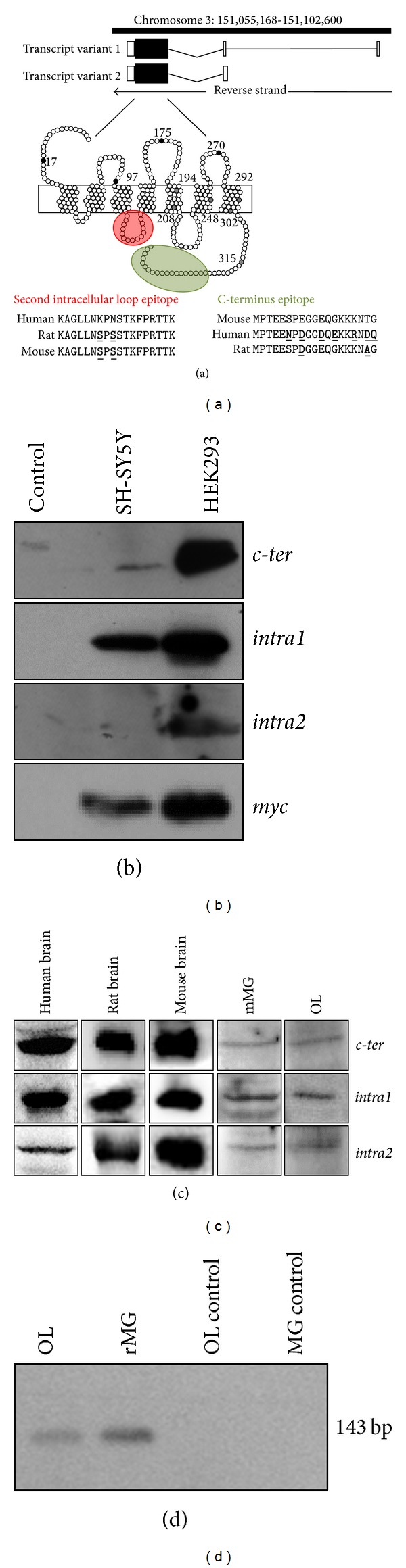
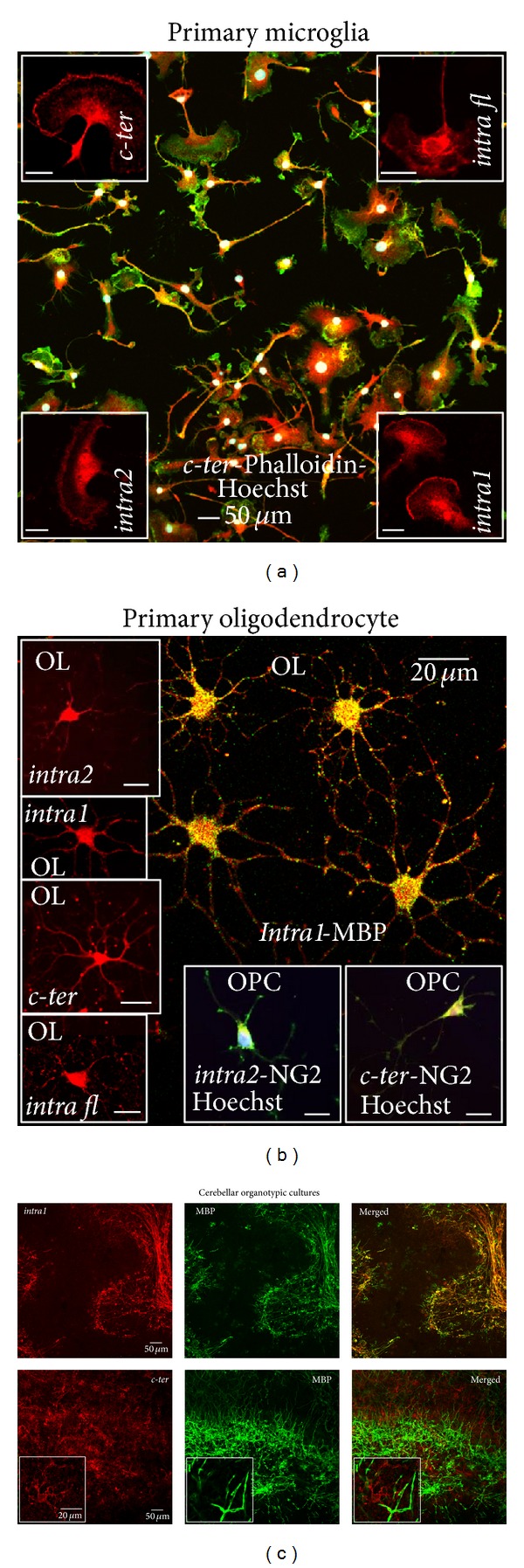
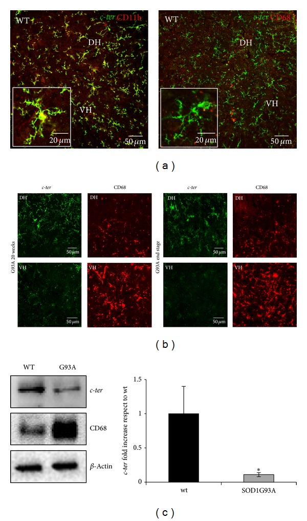
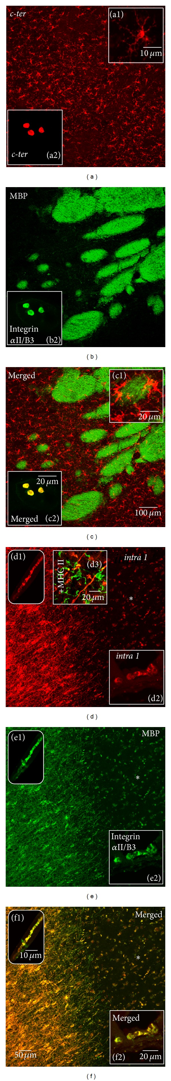
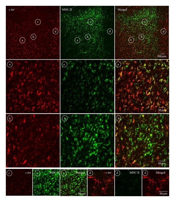
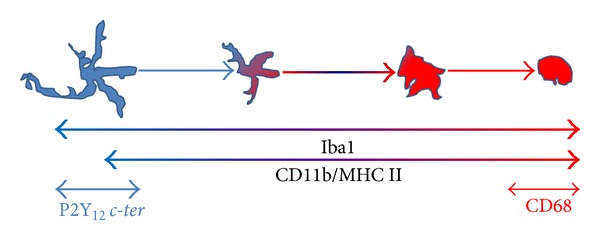
In the CNS, neuroinflammation occurring during pathologies as amyotrophic lateral sclerosis (ALS) and multiple sclerosis (MS) is the consequence of an intricate interplay orchestrated by various cell phenotypes. Among the molecular cues having a role in this process, extracellular nucleotides are responsible for intercellular communication and propagation of inflammatory stimuli. This occurs by binding to several receptor subtypes, defined P2X/P2Y, which are widespread in different tissues and simultaneously localized on multiple cells. For instance, the metabotropic P2Y12 subtype is found in the CNS on microglia, affecting activation and chemotaxis, on oligodendrocytes, possessing a hypothesized role in myelination, and on astrocytes. By comparative analysis, we have established here that P2Y12 receptor immunolabelled by antibodies against C-terminus or second intracellular loop, is, respectively, distributed and modulated under neuroinflammatory conditions on ramified microglia or myelinated fibers, in primary organotypic cerebellar cultures, tissue slices from rat striatum and cerebellum, spinal cord sections from symptomatic/end stage SOD1-G93A ALS mice, and finally autoptic cortical tissue from progressive MS donors. We suggest that modulation of P2Y12 expression might play a dual role as analytic marker of branched/surveillant microglia and demyelinating lesions, thus potentially acquiring a predictive value under neuroinflammatory conditions as those found in ALS and MS.


Figures

Figure 1
Figure 1
P2Y12 antibodies validation and RT-PCR analysis. (a) Scheme of human P2RY12 gene location, transcript variants [29, 30], and protein structure, with amino acid epitopes recognized by the used antibodies (Table 1) and highlighted in color (intra1, intra2, and intra fl, red circle; c-ter, green oval). Species conservation for each epitope was calculated by using BLAT tool of UCSC genome browser [31]. (b) Total protein extracts from SH-SY5Y or HEK293 cells expressing Myc-tagged P2Y12 receptor were subjected to Western blot analysis with the indicated antibodies. (c) Total protein extracted from human, rat and mouse brain, from primary mouse microglia (mMG) and rat oligodendrocyte (OL) cultures were subjected to Western blot analysis with the indicated antibodies. For intra2 antibodies,  lots AN01/02/04/0502/0602 were used. (d) RT-PCR using primers specific for P2Y12 mRNA was performed on total RNA from rat microglia (rMG) and OL. Control lanes show RT-PCR performed without reverse transcriptase enzyme.
Figure 2
Figure 2
P2Y12 receptor in dissociated and organotypic primary cultures. (a) Mouse primary cortical microglia were subjected to immunofluorescence and confocal analysis with phalloidin (green, merged field) and P2Y12 receptor antibodies (red, insets and merged) and Hoechst (white, merged). Scale bars in insets: 20 μm. (b) Double immunofluorescence and confocal analysis of primary rat mature (OL) and precursor (OPC) oligodendrocytes was performed with antibodies for P2Y12 receptor, MBP, NG2 (Table 1). For intra2 antibodies, lots AN01/02/04/0502/0602 were used. (c) Rat cerebellar organotypic cultures were analyzed by double immunofluorescence and confocal microscopy for intra1 (red) and c-ter (red), highlighting different structures (see also insets), and MBP (green).
Figure 3
Figure 3
P2Y12 receptor in rat brain tissue. Double immunofluorescence and confocal analysis was performed on sections from rat cerebellum (panels (a), (b), (c), (g), (h), and (i)) and striatum (panels (d), (e), (f), (j), (k), and (l)) with intra1, intra2-lots AN01/02/04, intra fl, c-ter (all red), and GFAP (green, inset b1), CD11b (green, insets c1, f1, h1; yellow merged, inset i1; green, panel (k); merged, panel (l)), MBP (yellow merged, inset c2; green, inset e1; green, panels (h) and (i); green, inset j1) antibodies.
Figure 4
Figure 4
Temporal and regional pattern of P2Y12 expression in SOD1-G93A ALS spinal microglia. (a) Double immunofluorescence and confocal analysis on lumbar spinal cord sections (L3–L5) of wild-type (WT) mice was performed with c-ter antibody (green and yellow, merged and insets), CD11b (left panel, yellow, merged and inset), and CD68 (right panel, red, merged and inset), in both dorsal (DH) and ventral (VH) horns of spinal cord. (b) Double immunofluorescence and confocal analysis on SOD1-G93A lumbar spinal cord sections (L3–L5) at two different stages of ALS disease, that is, 20 weeks, and end stage, was performed with c-ter (green) and CD68 (red) antibodies. (c) Equal amount of total lumbar spinal cord lysates (L3–L5) from WT and SOD1-G93A (n = 4 for each group) were subjected to Western blotting and immunoreactions with c-ter and CD68 antibodies; anti-β-actin was used for protein normalization. Data represent means ± SEM. Statistical significance was calculated by Student's t-test, *P < 0.05.
Figure 5
Figure 5
P2Y12 receptor in human cortex. Sections from human healthy and SPMS frontal cortex were analyzed by double immunofluorescence and confocal microscopy for the immunoreactive markers c-ter (red, panels (a), (c); insets a1, a2, c1; yellow merged, inset c2), intra1 (red, panels (d), insets d1, d2, d3; yellow merged, panel f, insets f1, f2), MBP (green, panels (b), (c), (e), insets c1, e1; yellow merged, panel (f), inset f1), MHC II (green, inset d3), and integrin αII/β3 (green, insets b2, e2; yellow merged, insets c2, f2). The asterisks show decreased P2Y12 immunoreactivity in proximity to MS lesion.
Figure 6
Figure 6
Regional distribution of P2Y12 in proximity to MS lesions. Sections from SPMS frontal cortex were analyzed by double immunofluorescence and confocal microscopy for c-ter (red) and MHC II (green) immunoreactivity. In proximity to the demyelinating active cortical lesion expressing augmented positivity for MHC II, microglia gradually lose immunoreactivity for c-ter antibody. Microglia express differential immunoreactivity in the four chosen areas which are found inside (circled b-c) and around (circled a–d) a lesion.
Figure 7
Figure 7
Draw of microglial marker expression as a function of activation. Branched microglia are represented in blue and activated microglia in red. Iba1 [22], CD11b, and MHC II are mostly expressed in microglia throughout the different morphological states and their expression increases during activation (light blue to red color). P2Y12 c-ter (light blue) and CD68 (red) are expressed, respectively, in branched or roundish/activated microglia.

References

    1. Guérout N, Li X, Barnabé-Heider F. Cell fate control in the developing central nervous system. Experimental Cell Research. 2014;321(1):77–83. - PubMed
    1. Philips T, Robberecht W. Neuroinflammation in amyotrophic lateral sclerosis: role of glial activation in motor neuron disease. The Lancet Neurology. 2011;10(3):253–263. - PubMed
    1. Ellwardt E, Zipp F. Molecular mechanisms linking neuroinflammation and neurodegeneration in MS. Experimental Neurology. 2014 - PubMed
    1. Walsh JG, Muruve DA, Power C. Inflammasomes in the CNS. Nature Reviews Neuroscience. 2014;15(2):84–97. - PubMed
    1. di Virgilio F, Ceruti S, Bramanti P, Abbracchio MP. Purinergic signalling in inflammation of the central nervous system. Trends in Neurosciences. 2009;32(2):79–87. - PubMed

Publication types

Substances

LinkOut - more resources