

Age-related changes of the ocular surface: a hospital setting-based retrospective study
- PMID: 25180084
- PMCID: PMC4144156
- DOI: 10.1155/2014/532378
Age-related changes of the ocular surface: a hospital setting-based retrospective study
Abstract
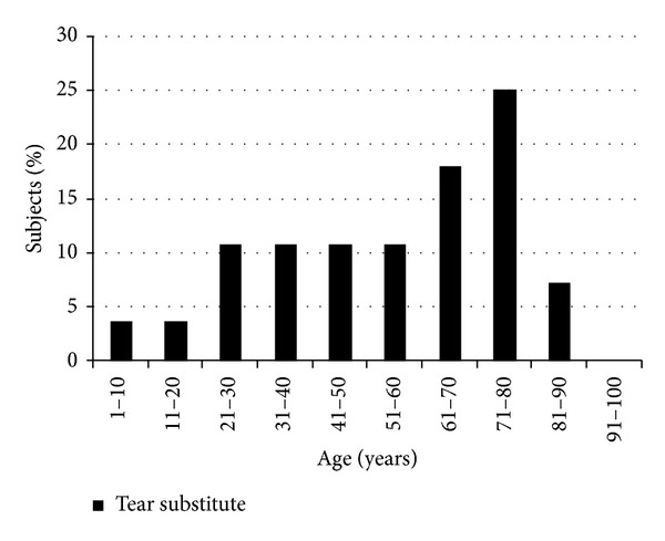
Purpose. To investigate the effects of age on the prevalence of ocular surface diseases (OSD), adherence to treatment, and recovery rates. Patients and Methods. Retrospective analysis of 3000 clinical records from a first-level general ophthalmology clinic. Patients with OSD were prospectively submitted a questionnaire to assess compliance and recovery rates. Results. OSD prevalence was 10.3%. Patients with OSD were significantly older than patients without it: 67.5 ± 20.3 versus 57.0 ± 22.0 years (P = 0.036). No significant difference in season distribution was shown. Dry eye disease (DED) represented 58% of OSD; its prevalence increased with age until 80 years old and suddenly decreased thereafter. Asymptomatic DED was 37%. Adherence to treatment in OSD was very high (94%); recovery rates were lower in patients aged 21-40 and 61-80 (resp., 65.5% and 77.8%) and this was associated with higher OSDI scores. Tear substitutes represented 50% of all prescribed medications; their use increased with age. Discussion. In a "real-life" low-tech setting, OSD showed a prevalence of 10.3%. DED was the most prevalent disease, and it was asymptomatic in more than 1/3 of cases.
Figures


References
-
- Lemp MA, Baudouin C, Baum J, et al. The definition and classification of dry eye disease: report of the definition and classification subcommittee of the international Dry Eye WorkShop (2007) Ocular Surface. 2007;5(2):75–92. - PubMed
-
- Gipson IK. Age-related changes and diseases of the ocular surface and cornea. Investigative Ophthalmology & Visual Science. 2013;54(14):48–53. - PubMed
-
- Wei A, Hong J, Sun X, Xu J. Evaluation of age-related changes in human palpebral conjunctiva and meibomian glands by in vivo confocal microscopy. Cornea. 2011;30(9):1007–1012. - PubMed
-
- De Cilla S, Fogagnolo P, Sacchi M, et al. Corneal involvement in uneventful cataract surgery: an in vivo confocal microscopy study. Ophthalmologica. 2014;231(2):103–110. - PubMed
-
- de Cillà S, Ranno S, Carini E, et al. Corneal subbasal nerves changes in patients with diabetic retinopathy: an in vivo confocal study. Investigative Ophthalmology and Visual Science. 2009;50(11):5155–5158. - PubMed
LinkOut - more resources
Full Text Sources
Other Literature Sources
