Identification of paralogous life-cycle stage specific cytoskeletal proteins in the parasite Trypanosoma brucei
- PMID: 25180513
- PMCID: PMC4152294
- DOI: 10.1371/journal.pone.0106777
Identification of paralogous life-cycle stage specific cytoskeletal proteins in the parasite Trypanosoma brucei
Abstract
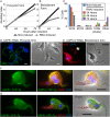
The life cycle of the African trypanosome Trypanosoma brucei, is characterised by a transition between insect and mammalian hosts representing very different environments that present the parasite with very different challenges. These challenges are met by the expression of life-cycle stage-specific cohorts of proteins, which function in systems such as metabolism and immune evasion. These life-cycle transitions are also accompanied by morphological rearrangements orchestrated by microtubule dynamics and associated proteins of the subpellicular microtubule array. Here we employed a gel-based comparative proteomic technique, Difference Gel Electrophoresis, to identify cytoskeletal proteins that are expressed differentially in mammalian infective and insect form trypanosomes. From this analysis we identified a pair of novel, paralogous proteins, one of which is expressed in the procyclic form and the other in the bloodstream form. We show that these proteins, CAP51 and CAP51V, localise to the subpellicular corset of microtubules and are essential for correct organisation of the cytoskeleton and successful cytokinesis in their respective life cycle stages. We demonstrate for the first time redundancy of function between life-cycle stage specific paralogous sets in the cytoskeleton and reveal modification of cytoskeletal components in situ prior to their removal during differentiation from the bloodstream form to the insect form. These specific results emphasise a more generic concept that the trypanosome genome encodes a cohort of cytoskeletal components that are present in at least two forms with life-cycle stage-specific expression.
Conflict of interest statement
Figures


Similar articles
-
Functional Analyses of Cytokinesis Regulators in Bloodstream Stage Trypanosoma brucei Parasites Identify Functions and Regulations Specific to the Life Cycle Stage.mSphere. 2019 May 1;4(3):e00199-19. doi: 10.1128/mSphere.00199-19. mSphere. 2019. PMID: 31043517 Free PMC article.
-
Cytokinesis in Trypanosoma brucei differs between bloodstream and tsetse trypomastigote forms: implications for microtubule-based morphogenesis and mutant analysis.Mol Microbiol. 2013 Dec;90(6):1339-55. doi: 10.1111/mmi.12436. Epub 2013 Nov 15. Mol Microbiol. 2013. PMID: 24164479 Free PMC article.
-
Cytokinesis in bloodstream stage Trypanosoma brucei requires a family of katanins and spastin.PLoS One. 2012;7(1):e30367. doi: 10.1371/journal.pone.0030367. Epub 2012 Jan 18. PLoS One. 2012. PMID: 22279588 Free PMC article.
-
Proteomics and the Trypanosoma brucei cytoskeleton: advances and opportunities.Parasitology. 2012 Aug;139(9):1168-77. doi: 10.1017/S0031182012000443. Epub 2012 Apr 4. Parasitology. 2012. PMID: 22475638 Review.
-
The cell biology of Trypanosoma brucei differentiation.Curr Opin Microbiol. 2007 Dec;10(6):539-46. doi: 10.1016/j.mib.2007.09.014. Epub 2007 Nov 9. Curr Opin Microbiol. 2007. PMID: 17997129 Free PMC article. Review.
Cited by
-
Novel Cytoskeleton-Associated Proteins in Trypanosoma brucei Are Essential for Cell Morphogenesis and Cytokinesis.Microorganisms. 2021 Oct 27;9(11):2234. doi: 10.3390/microorganisms9112234. Microorganisms. 2021. PMID: 34835360 Free PMC article.
-
Genome-scale RNA interference profiling of Trypanosoma brucei cell cycle progression defects.Nat Commun. 2022 Sep 10;13(1):5326. doi: 10.1038/s41467-022-33109-y. Nat Commun. 2022. PMID: 36088375 Free PMC article.
-
Post-transcriptional reprogramming by thousands of mRNA untranslated regions in trypanosomes.Nat Commun. 2024 Sep 16;15(1):8113. doi: 10.1038/s41467-024-52432-0. Nat Commun. 2024. PMID: 39285175 Free PMC article.
-
Genome-wide subcellular protein map for the flagellate parasite Trypanosoma brucei.Nat Microbiol. 2023 Mar;8(3):533-547. doi: 10.1038/s41564-022-01295-6. Epub 2023 Feb 20. Nat Microbiol. 2023. PMID: 36804636 Free PMC article.
-
Distinct Phenotypes Caused by Mutation of MSH2 in Trypanosome Insect and Mammalian Life Cycle Forms Are Associated with Parasite Adaptation to Oxidative Stress.PLoS Negl Trop Dis. 2015 Jun 17;9(6):e0003870. doi: 10.1371/journal.pntd.0003870. eCollection 2015 Jun. PLoS Negl Trop Dis. 2015. PMID: 26083967 Free PMC article.
References
-
- Vickerman K (1985) Developmental cycles and biology of pathogenic trypanosomes. Br. Med. Bull. 41: 105–114. - PubMed
-
- Sharma R, Peacock L, Gluenz E, Gull K, Gibson W, et al. (2008) Asymmetric cell division as a route to reduction in cell length and change in cell morphology in trypanosomes. Protist 159: 137–151. - PubMed
-
- Sherwin T, Gull K (1989) Visualization of detyrosination along single microtubules reveals novel mechanisms of assembly during cytoskeletal duplication in trypanosomes. Cell 57: 211–221. - PubMed
Publication types
MeSH terms
Substances
Grants and funding
LinkOut - more resources
Full Text Sources
Other Literature Sources
Miscellaneous

