. 2014 Sep 2;9(9):e106535.
doi: 10.1371/journal.pone.0106535. eCollection 2014.

Inhibition of West Nile virus by calbindin-D28k

Affiliations

Inhibition of West Nile virus by calbindin-D28k

Venkatraman Siddharthan et al. PLoS One. .

Abstract

Evidence indicates that West Nile virus (WNV) employs Ca(2+) influx for its replication. Moreover, calcium buffer proteins, such as calbindin D28k (CB-D28k), may play an important role mitigating cellular destruction due to disease processes, and more specifically, in some neurological diseases. We addressed the hypothesis that CB-D28k inhibits WNV replication in cell culture and infected rodents. WNV envelope immunoreactivity (ir) was not readily co-localized with CB-D28k ir in WNV-infected Vero 76 or motor neuron-like NSC34 cells that were either stably or transiently transfected with plasmids coding for CB-D28k gene. This was confirmed in cultured cells fixed on glass coverslips and by flow cytometry. Moreover, WNV infectious titers were reduced in CB-D28k-transfected cells. As in cell culture studies, WNV env ir was not co-localized with CB-D28k ir in the cortex of an infected WNV hamster, or in the hippocampus of an infected mouse. Motor neurons in the spinal cord typically do not express CB-D28k and are susceptible to WNV infection. Yet, CB-D28k was detected in the surviving motor neurons after the initial phase of WNV infection in hamsters. These data suggested that induction of CB-D28k elicit a neuroprotective response to WNV infection.


Conflict of interest statement

Competing Interests: The authors have declared that no competing interests exist.

Figures

Figure 1
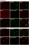
Figure 1. Reduced co-localization of WNV immunoreactivity (ir) with CB-D28k ir in (A) Vero 76 cells and (B) NSC34 cells transiently transfected with CB-D28k, vector control, or pGAG-GFP plasmids.
Cells at 70–80% confluence were infected with a multiplicity of infection (MOI) of 5. Two days after viral challenge, cells on glass coverslips were fixed and immunostained. Arrows show co-localization of WNV with gene products of transfected plasmid. The transfection control was pCAG-GFP – CAG strong synthetic promoter for mammalian expression of green fluorescent protein (GFP).
Figure 2
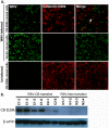
Figure 2. Reduced co-localization of WNV immunoreactivity (ir) with CB-D28k ir in NSC34 cells stably transfected with CB-D28k plasmid.
(A) Cells stably transfected with RSV promoter–calbindin D28K/neomycin resistance (RSV-CB) or RSV promoter–neomycin resistance (RSV-Neo) plasmids were infected with 5 multiplicity of infection (MOI) and immunostained 48 hours later. Arrow shows co-localization of WNV env ir with CB-D28k ir. (B) Western blot of CB-D28K in stably transfected NSC34 cloned cell lines. Clones C1–2 and N1–1 were used for WNV infection.
Figure 3
Figure 3. Quantitation of co-localization of CB-D28k ir with WNV env ir.
(A) Stable transfection. Cell culture mixture of 50% CB-D28k stably transfected NSC34 cells and 50% vector control stably transfected NSC34 cells at 70–80% confluence were infected with 5 MOI and immunostained 48 hr later. The analyzed images were obtained from 5 representative areas of the slides using confocal fluorescent microscope. From those images, using ZEN software, based on the intensity of the staining, WNV-infected and CB-D28K transfected cells were quantified and plotted. The area within the dotted-line box represent cells where some level of co-localization of CB-D28k ir and WNV env ir was detectable. If CB-D28k did not affect viral replication, there would be no statistical difference between 50% of the number inside the box compared with 50% of the number outside the box. Therefore, chi square statistical analysis was performed by comparing the number of cells co-localized inside the box with the number of cells outside the box. (B) Transient transfection. NSC34 cells were transiently transfected with CB-D28k expression plasmid, and seeded at 70% confluence on glass coverslips. The next day the cells were infected with 5 MOI and analyzed by flow cytometry (total of 73 cells analyzed). Chi square statistical analysis was performed by conservatively assuming at an infection of 5 MOI that at least 60% of the CB-D28k ir cells would be infected by WNV, and co-localized with WNV env ir cells, and comparing the numbers inside and outside the box. P≤0.001. (C) Frequency of distribution of CB-D28k ir in stably transfected NSC34 cells. A total of 260 cells in fields of 100% stably transfected cells through three rounds of cloning were individually measured for relative ir and plotted on a frequency distribution histogram. Y-axis is represented as the percent (%) of total frequency.
Figure 4
Figure 4. Reduced WNV replication in NSC34 cells expressing CB-D28k as detected by flow cytometry.
NSC34 cells stably transfected with (A) the vector control plasmid or (B) CB-D28k plasmid were infected with WN02 at 5 MOI one day after seeding 6-well plates at 70–80% confluence. After 1-hour of adsorption, cells were washed and incubated for 48 hours. Cells were detached with 0.1% trypsin in MEM without FBS, and double-immunostained for WNV env (green-Alexa fluor 488) and CB-D28k (red-Alexa fluor 568). Flow cytometry analysis was performed with 10,000 cells for each group. NSC34 cells stably transfected with the vector control were 87.9% (83.5%+4.4%) immunoreactive for WNV env, as compared to 36.2% (12.7%+23.5%) with cells expressing CB-D28k. Histogram overlays reveal distinct peaks of CB-D28k-positive and –negative cells (C), and WNV-positive and -negative cells (D).
Figure 5
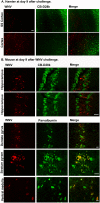
Figure 5. Absence of co-localization of CB-D28k ir with WNV env ir in (A) hamster, and (B) mouse neurological tissues. Rodents were infected s.c. with NY99 WNV.
When WNV-infected animals could not right themselves, necropsy was performed, and immune-stained tissues were inspected for areas containing WNV env ir, plus CB-D28k ir or parvalbumin ir. SS Cortex: somatosensory cortex, Cortex: cerebral cortex, pyram: pyramidale. Bars = 50 µm.
Figure 6
Figure 6. Induction of CB-D28k in spinal cord motor neurons by infection of hamsters by WNV.
Hamsters were injected subcutaneously with NY99 WNV or sham (uninfected cell homogenate), and necropsied when WNV-infected animals could not right themselves. The cervical spinal cord was processed for immunohistochemistry (left). Intensity of CB-D28k was plotted from tissues in WNV- and sham-infected hamsters (right). Each dot represents individual cells from 4 different hamsters. ***P≤0.01 using t test.
Figure 7
Figure 7. Induction of CB-D28K in the spinal cords of hamsters by infection with WNV.
(A) Immunoperoxidase staining of CB-D28k in WNV- and sham-infected hamsters. Arrows: increased staining in motor neurons. (B) Hamsters were infected subcutaneously with WNV or sham. At 6, 8, 11, 16, and 21 days post-viral injection (dpi), subsets of hamsters were euthanized for collection of spinal cord tissues that were processed by western blot analysis for detection of CB-D28K. (C) Quantification of CB-D28k by western blot. Fold-increases were based on comparison to the sham-infected data. **P≤0.01, ***P≤0.001 compared to sham-infected data using ANOVA with Neuman-Keuls multiple comparison test.

References

    1. Schwaller B (2010) Cytosolic Ca2+ buffers. Cold Spring Harbor perspectives in biology 2: a004051. - PMC - PubMed
    1. Zhou Y, Frey TK, Yang JJ (2009) Viral calciomics: interplays between Ca2+ and virus. Cell calcium 46: 1–17. - PMC - PubMed
    1. Scherbik SV, Brinton MA (2010) Virus-induced Ca2+ influx extends survival of west nile virus-infected cells. Journal of Virology 84: 8721–8731. - PMC - PubMed
    1. Christakos S, Barletta F, Huening M, Dhawan P, Liu Y, et al. (2003) Vitamin D target proteins: function and regulation. Journal of cellular biochemistry 88: 238–244. - PubMed
    1. Mattson MP, Rychlik B, Chu C, Christakos S (1991) Evidence for calcium-reducing and excito-protective roles for the calcium-binding protein calbindin-D28k in cultured hippocampal neurons. Neuron 6: 41–51. - PubMed

Publication types

MeSH terms