Review
. 2014 Nov 14;12(42):8367-78.
doi: 10.1039/c4ob01063a.

Mechanisms for enzymatic cleavage of the N-glycosidic bond in DNA

Affiliations
Review

Mechanisms for enzymatic cleavage of the N-glycosidic bond in DNA

Alexander C Drohat et al. Org Biomol Chem. .

Abstract

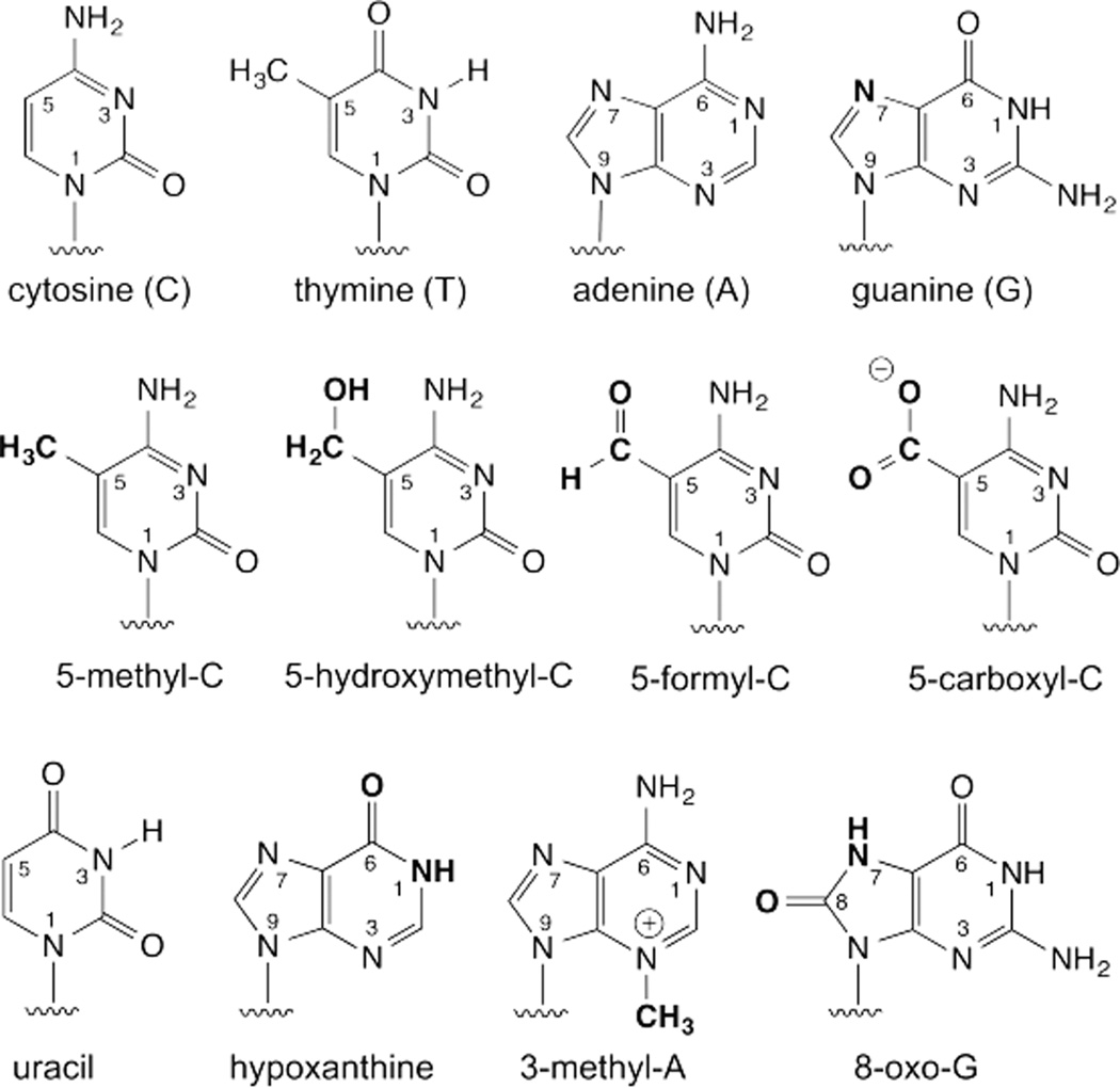
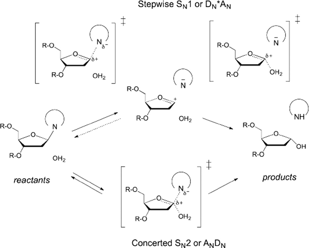
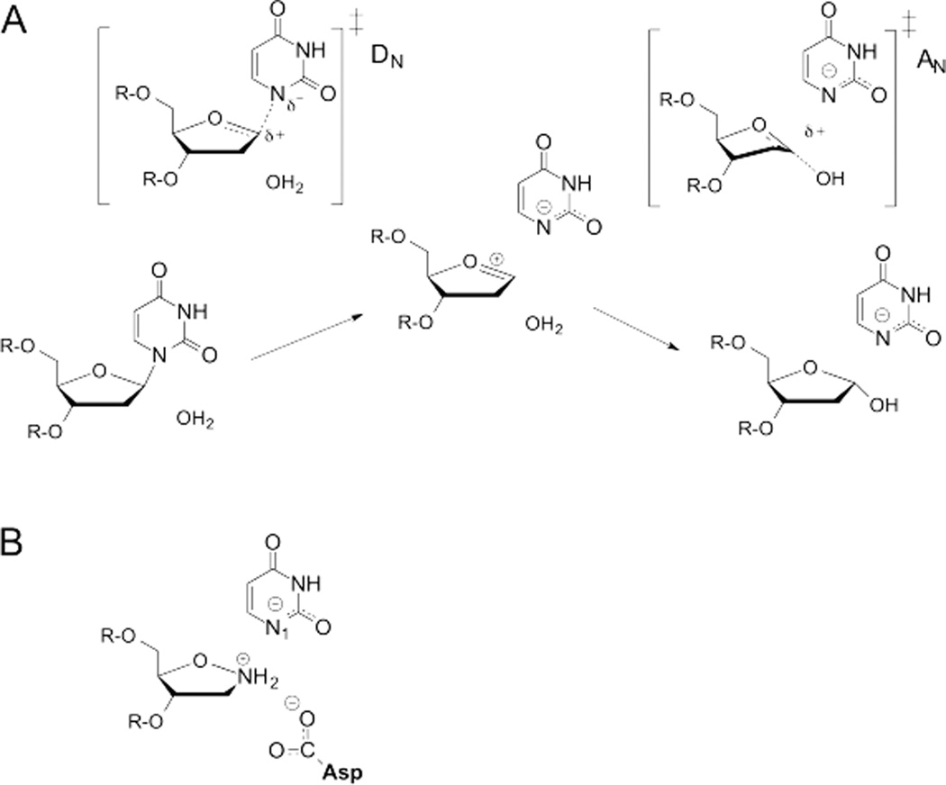
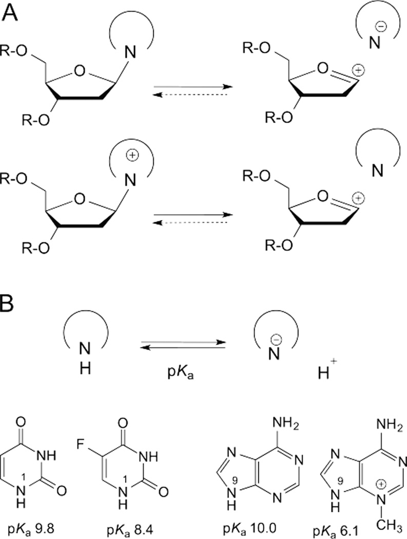
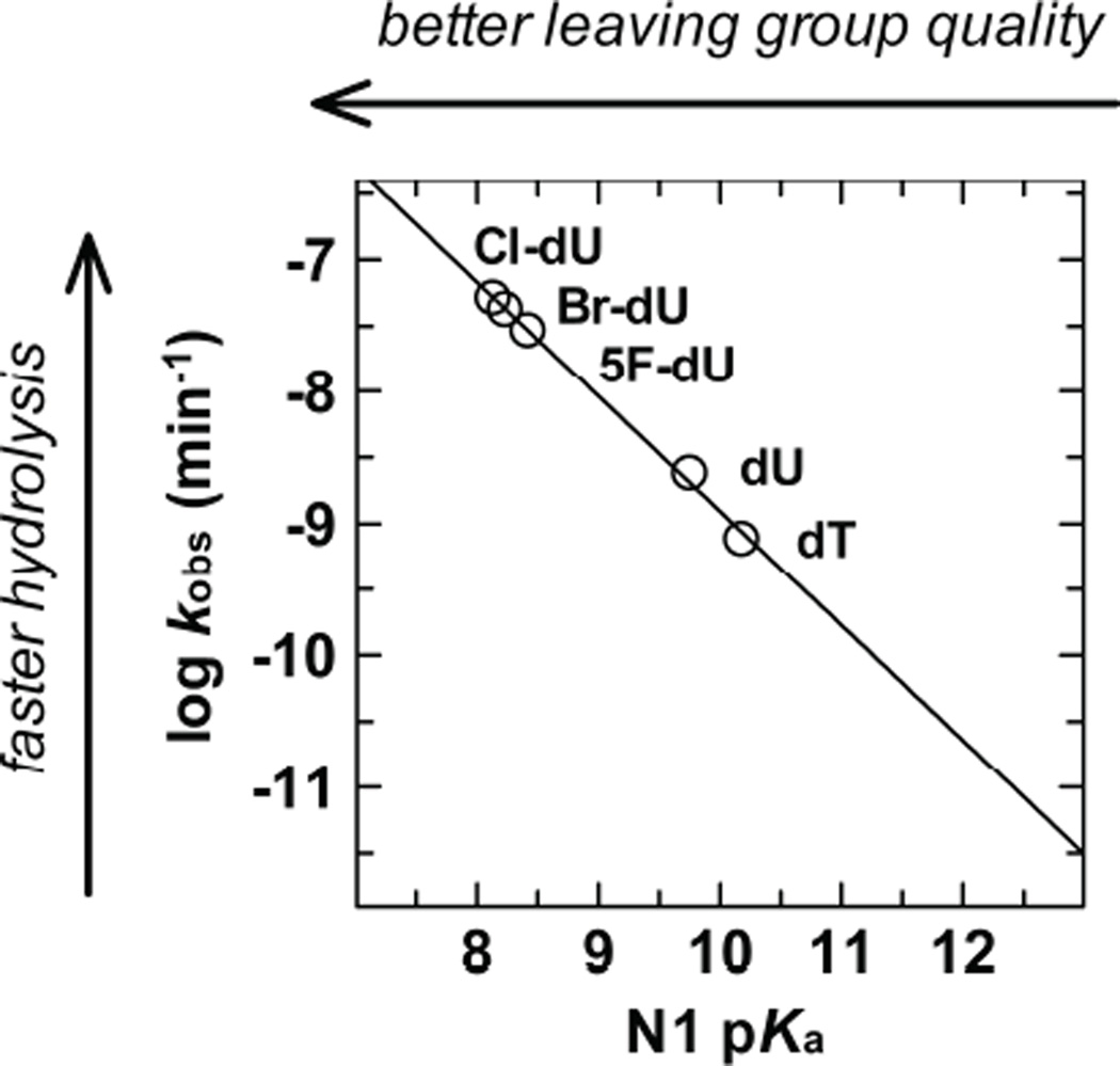
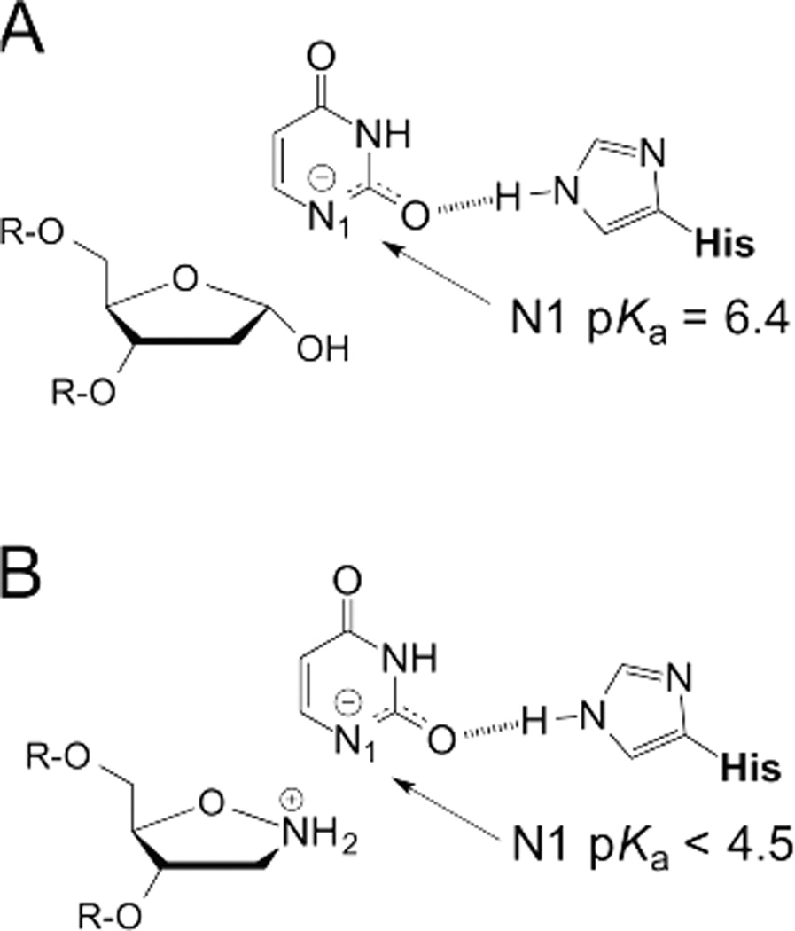
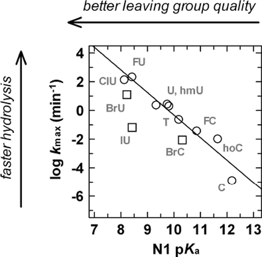
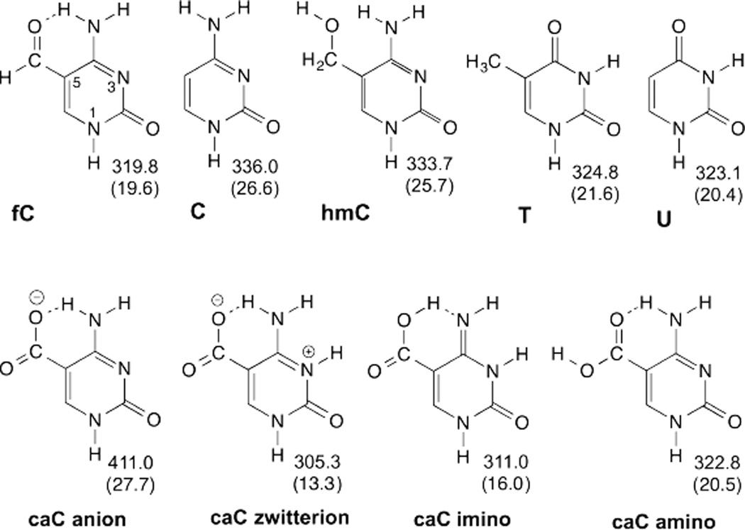
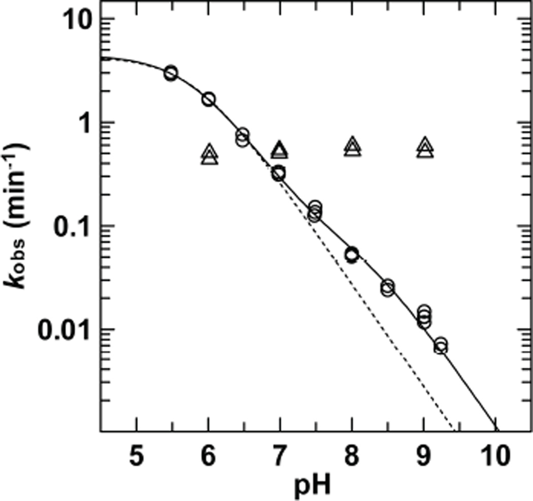
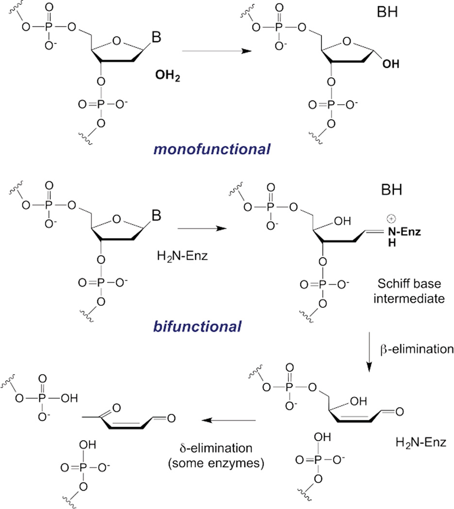
DNA glycosylases remove damaged or enzymatically modified nucleobases from DNA, thereby initiating the base excision repair (BER) pathway, which is found in all forms of life. These ubiquitous enzymes promote genomic integrity by initiating repair of mutagenic and/or cytotoxic lesions that arise continuously due to alkylation, deamination, or oxidation of the normal bases in DNA. Glycosylases also perform essential roles in epigenetic regulation of gene expression, by targeting enzymatically-modified forms of the canonical DNA bases. Monofunctional DNA glycosylases hydrolyze the N-glycosidic bond to liberate the target base, while bifunctional glycosylases mediate glycosyl transfer using an amine group of the enzyme, generating a Schiff base intermediate that facilitates their second activity, cleavage of the DNA backbone. Here we review recent advances in understanding the chemical mechanism of monofunctional DNA glycosylases, with an emphasis on how the reactions are influenced by the properties of the nucleobase leaving-group, the moiety that varies across the vast range of substrates targeted by these enzymes.


Figures

Fig. 1
Fig. 1
The four canonical bases in DNA (top), four enzymatically-generated forms of cytosine found in vertebrates (middle), and four examples of damaged bases (bottom) from among the many dozens that arise in DNA.
Fig. 2
Fig. 2
Two limiting mechanisms for N-glycosidic bond hydrolysis in (deoxy)ribonucleosides. The stepwise reaction (top) has two transition states, for departure of the leaving group (DN) and nucleophile addition (AN).
Fig. 3
Fig. 3
UNG-catalyzed excision of uracil from DNA. (A) Transition-state analysis and computational studies indicate that UNG follows a stepwise mechanism with irreversible C-N bond cleavage. NMR studies show that the uracil anion is stabilized in the ternary product complex. (B) The uracil anion and a conserved Asp side chain interact with the 1-aza-2'-deoxyribose cation, suggesting similar stabilization of the oxacarbenium ion in the chemical transition state(s).
Fig. 4
Fig. 4
Leaving group quality of the nucleobase depends on the acidity (pKa) of its glycosidic nitrogen. (A) In the stepwise reactions for glycosidic bond hydrolysis, a neutral base departs as an anion and a cationic base departs as a neutral species. (B) Acidity of the glycosidic nitrogen for a given base depends on the stability of the N-deprotonated species (conjugate base). The glycosidic nitrogen is N1 for pyrimidines, N9 for purines. Acidity (pKa) is shown for U, 5FU, A, and 3-methyl-A.
Fig. 5
Fig. 5
Damaged forms of adenine and guanine. AAG can remove Hx, εA, and 7meG but not 7-deaza-Hx.
Fig. 6
Fig. 6
Resonance structures of the 8-oxoguanine anion.
Fig. 7
Fig. 7
2’-deoxyxanthosine (dX) ionizes at N3 with a pKa of about 5.7 (based on xanthosine) and is predominantly anionic at pH 7.4. Hydrolysis of neutral dX results in departure of the xanthine (X) monoanion, which should be a good leaving group, given that N9 is acidic for X relative to other purines, due perhaps to charge delocalization (N7, O6).
Fig. 8
Fig. 8
(A) Hydrolysis of dU, dT, and dU analogs is not acid catalyzed at neutral pH, for enzymatic or non-enzymatic reactions, because the carbonyl oxygens are highly acidic (pKa < −3), so the base departs as an anion. (B) Resonance stabilization of the uracil anion involves the two carbonyl oxygens (O2, O4).
Fig. 9
Fig. 9
Brønsted-type linear free energy relationship (LFER) for non-enzymatic hydrolysis of 5-substituted deoxyuridines. The dependence of log kobs on N1 pKa (acidity of the leaving-group nitrogen) for hydrolysis of dT, dU, 5-F-dU, 5-Br-dU, and 5-Cl-dU gives a slope of βlg = −0.87 ± 0.03. The kobs values were extrapolated to 22 °C using values at higher temperatures,, as described. For 5-Br-dU, a corrected N1 pKa of 8.24 was used rather than the previously reported 8.49.
Fig. 10
Fig. 10
Enzymatic stabilization of anionic leaving group. UNG provides a strong hydrogen bond to stabilize the uracil anion in the product complex, as indicated by a reduced N1 pKa of 6.4 relative to that of 9.8 for uracil in solution. (B) The uracil N1 pKa is even further reduced in a ternary complex with UNG and DNA that contains a mimic of the cationic oxacarbenium ion.
Fig. 11
Fig. 11
Brønsted-type LFER for enzymatic (TDG) hydrolysis of 5-substituted dU and dC nucleotides in DNA as reported by Bennett et al. The dependence of log kobs on N1 pKa for the base gives βlg = −1.6 ± 0.2; included in the fitting are U, T, FU, ClU, hoU, hmU, FC, hoC, and C (O). Data for BrU, IU, and BrC (□) were not included in the fitting, because kmax values suggested limited access of these bases to the TDG active site.
Fig. 12
Fig. 12
Calculated N1 acidity for pyrimidines are given as the free energy (ΔG, kcal mol−1) of deprotonation in the gas phase and bulk solution (parenthetical values). Lower values indicate greater N1 acidity and better leaving-group quality.
Fig. 13
Fig. 13
pH dependence of TDG activity for G.fC (Δ) and G.caC (O) substrates. The G.caC data is best fitted to a model for ionization of one essential protonated group (pKa1 = 5.75 ± 0.03) and a second group (pKa2 = 8.2 ± 0.7) that is not essential but confers higher activity when deprotonated (solid line). Fitting is poor for a model with a single essential protonated group (dotted line).
Scheme 1
Scheme 1
N-glycosidic bond hydrolysis in DNA by monofunctional and bifunctional DNA glycosylases

References

    1. Lindahl T. Nature. 1993;362:709–715. - PubMed
    1. Hitomi K, Iwai S, Tainer JA. DNA Repair (Amst) 2007;6:410–428. - PubMed
    1. Brooks SC, Adhikary S, Rubinson EH, Eichman BF. Biochim. Biophys. Acta. 2013;1834:247–271. - PMC - PubMed
    1. Stivers JT, Jiang YL. Chem. Rev. 2003;103:2729–2759. - PubMed
    1. Berti PJ, McCann JA. Chem. Rev. 2006;106:506–555. - PubMed

LinkOut - more resources