. 2014 Sep 17;136(37):13053-8.
doi: 10.1021/ja507169f. Epub 2014 Sep 9.

Unraveling the mystery of ATP hydrolysis in actin filaments

Affiliations

Unraveling the mystery of ATP hydrolysis in actin filaments

Martin McCullagh et al. J Am Chem Soc. .

Abstract

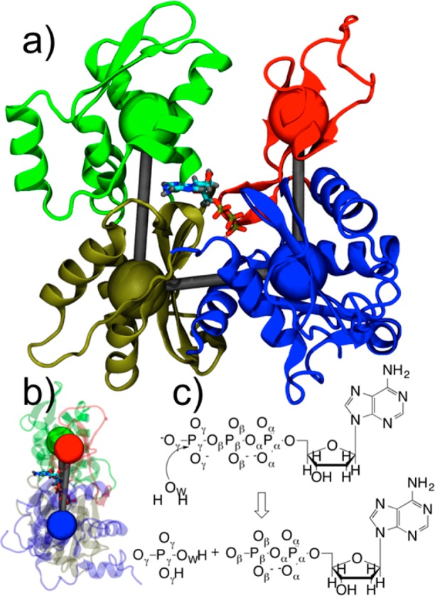
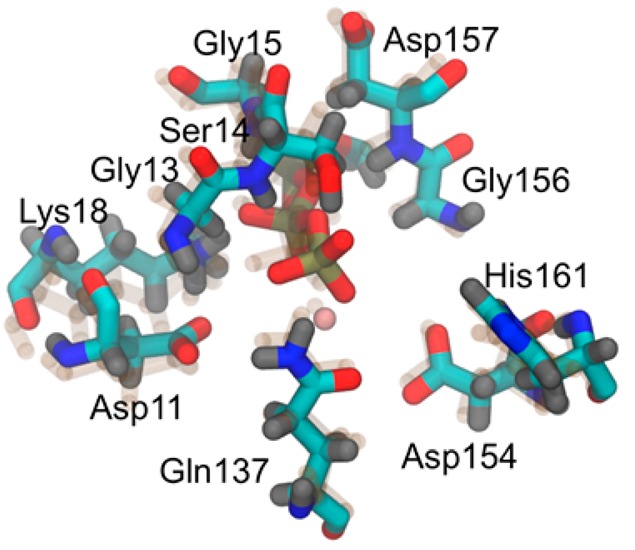
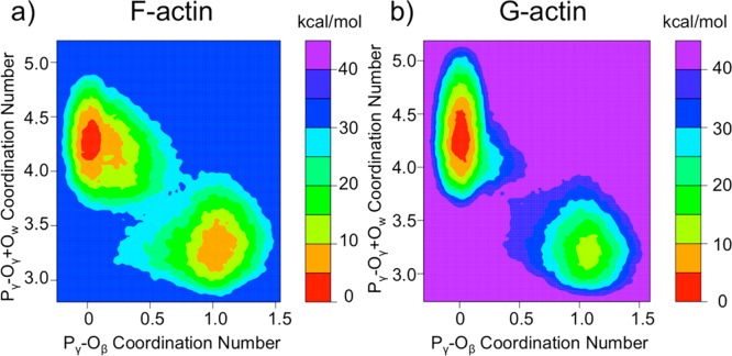
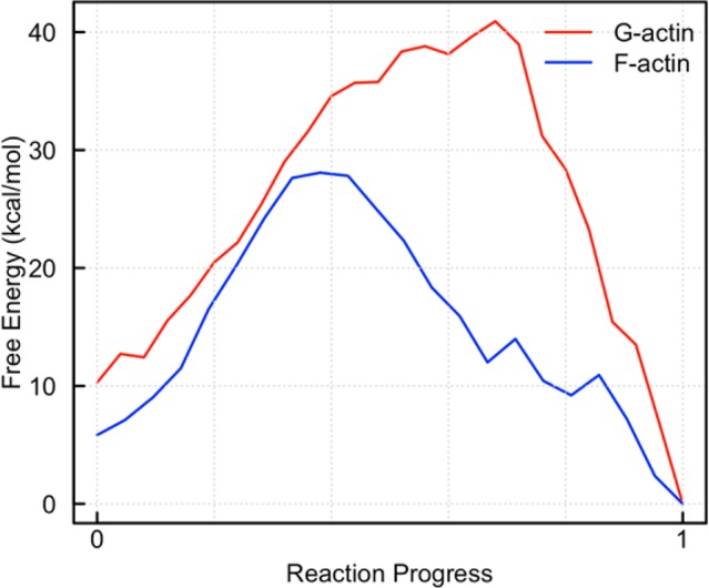
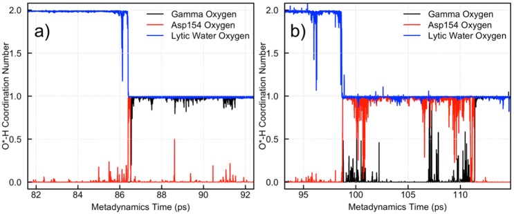
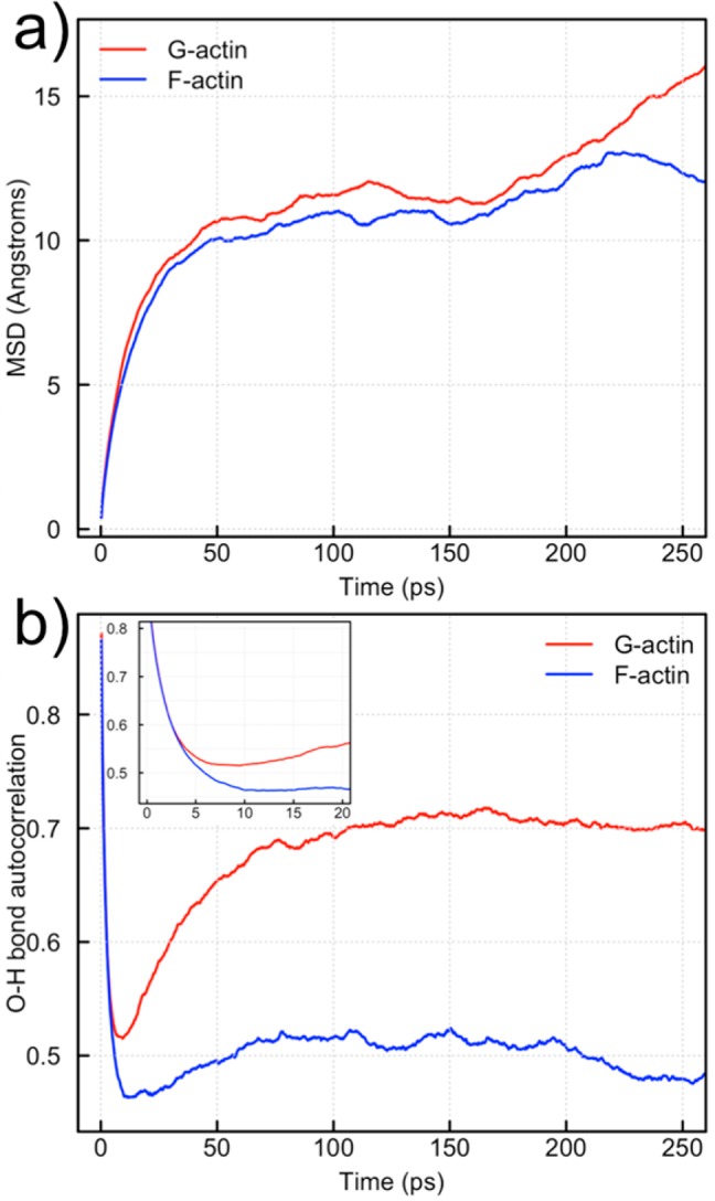
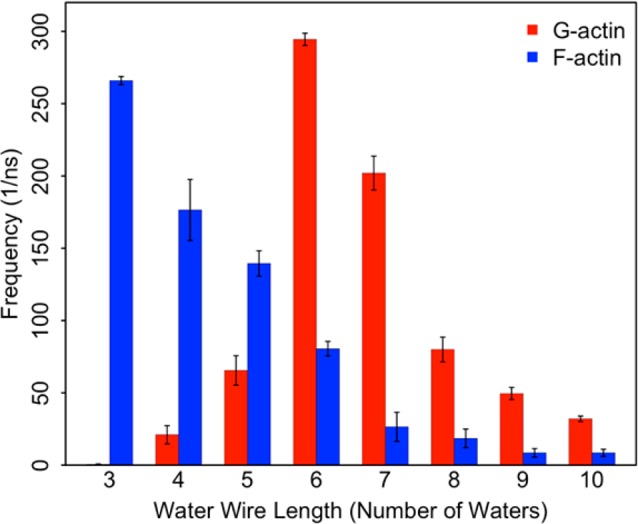
Actin performs its myriad cellular functions by the growth and disassembly of its filamentous form. The hydrolysis of ATP in the actin filament has been shown to modulate properties of the filament, thus making it a pivotal regulator of the actin life cycle. Actin has evolved to selectively hydrolyze ATP in the filamentous form, F-actin, with an experimentally observed rate increase over the monomeric form, G-actin, of 4.3 × 10(4). The cause of this dramatic increase in rate is investigated in this paper using extensive QM/MM simulations of both G- and F-actin. To compute the free energy of hydrolysis in both systems, metadynamics is employed along two collective variables chosen to describe the reaction coordinates of hydrolysis. F-actin is modeled as a monomer with restraints applied to coarse-grained variables enforced to keep it in a filament-like conformation. The simulations reveal a barrier height reduction for ATP hydrolysis in F-actin as compared to G-actin of 8 ± 1 kcal/mol, in good agreement with the experimentally measured barrier height reduction of 7 ± 1 kcal/mol. The barrier height reduction is influenced by an enhanced rotational diffusion of water in F-actin as compared to G-actin and shorter water wires between Asp154 and the nucleophilic water in F-actin, leading to more rapid proton transport.


Figures

Figure 1
Figure 1
(a) Structure of G-actin with four subdomains colored differently and coarse-grained variables depicted as colored spheres. Subdomain (SD) 1 is in blue, SD2 in red, SD3 in gold, and SD4 in green. ATP and magnesium ion are also depicted. (b) Side view of actin with SD2–SD1–SD3–SD4 dihedral angle in flat, F-actin conformation. (c) ATP hydrolysis reaction with atom labels.
Figure 2
Figure 2
Depiction of the nucleotide binding cleft of G-actin and F-actin (transparent). The 10 amino acids in the QM region are all labeled.
Figure 3
Figure 3
Two-dimensional free energy surfaces calculated for ATP hydrolysis in (a) F-actin and (b) G-actin. These were computed using metadynamics in QM/MM simulations. The dark blue region in panel a and the purple region in panel b represent unsampled areas.
Figure 4
Figure 4
Minimum free energy path from 2D free energy surfaces for G- and F-actin. The x-axis denotes reaction progress, with “0” being ATP and “1” being ADP+Pi. The position along the x-axis at which the transition state occurs is meaningless, but the barrier heights and relative energies are meaningful. The error analysis for metadynamics is not straightforward, but the error at each position is on the order of the hill height, which is 1.0 kcal/mol.
Figure 5
Figure 5
Monitoring of proton transport in (a) F-actin and (b) G-actin during ATP hydrolysis.
Figure 6
Figure 6
Quantification of water dynamics in the nucleotide binding pockets of G- and F-actin. (a) The mean squared displacement (MSD) of waters within 6 Å of the Pγ atom in both G- and F-actin. (b) The O–H bond autocorrelation for waters within 6 Å of the Pγ atom for both G- and F-actin. The inset is a blow-up of the time scales relevant to proton transport.
Figure 7
Figure 7
A histogram of the water wire length observed between a putative lytic water and Asp154 in both G- and F-actin. The error bars are computed using bootstrapping.
Figure 8
Figure 8
Snapshots of water wires leading to Asp154 from a putative lytic water from classical MD simulations of (a) F-actin and (b) G-actin. The F-actin snapshot (a) has a three-water wire, and the G-actin snapshot (b) has a six-water wire.

References

    1. Carlier M. F. In Actin Based Motility: Cellular, Molecular and Physical Aspects; Carlier M. F., Ed.; Springer: Dordrecht, Netherlands, 2010.
    1. Rould M. A.; Wan Q.; Joel P. B.; Lowey S.; Trybus K. M. J. Biol. Chem. 2006, 281, 31909. - PubMed
    1. Pollard T. D. J. Cell Biol. 1986, 103, 2747. - PMC - PubMed
    1. Blanchoin L.; Pollard T. D. Biochemistry 2002, 41, 597. - PubMed
    1. Kabsch W.; Mannherz H. G.; Suck D.; Pai E. F.; Holmes K. C. Nature 1990, 347, 37. - PubMed

Publication types

LinkOut - more resources