. 2014 Sep 2;9(9):e105834.
doi: 10.1371/journal.pone.0105834. eCollection 2014.

Cloning and functional analysis of three diacylglycerol acyltransferase genes from peanut (Arachis hypogaea L.)

Affiliations

Cloning and functional analysis of three diacylglycerol acyltransferase genes from peanut (Arachis hypogaea L.)

Xiaoyuan Chi et al. PLoS One. .

Abstract

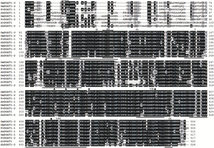
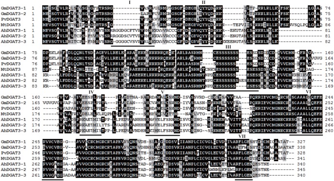
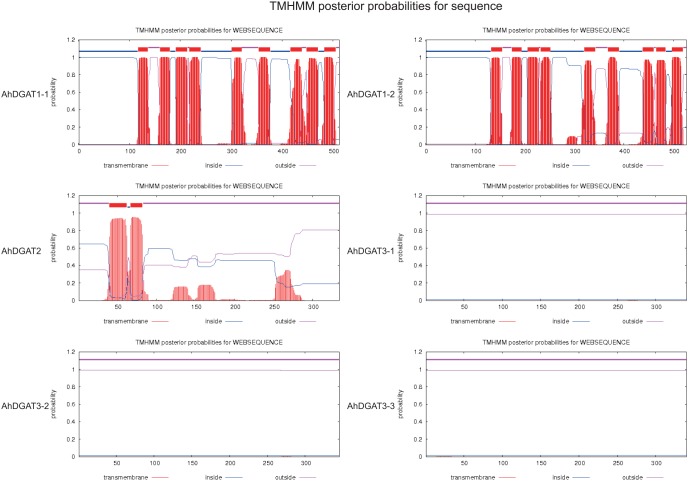
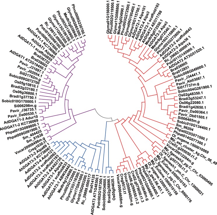
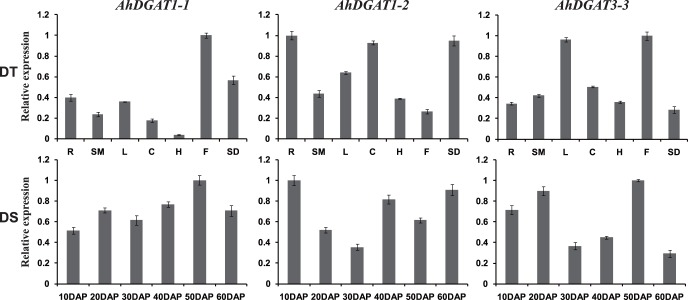
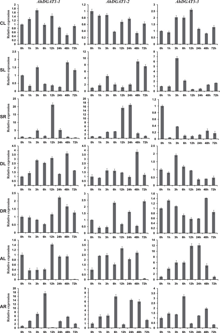
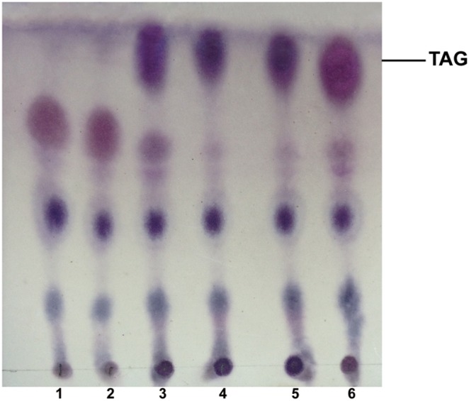
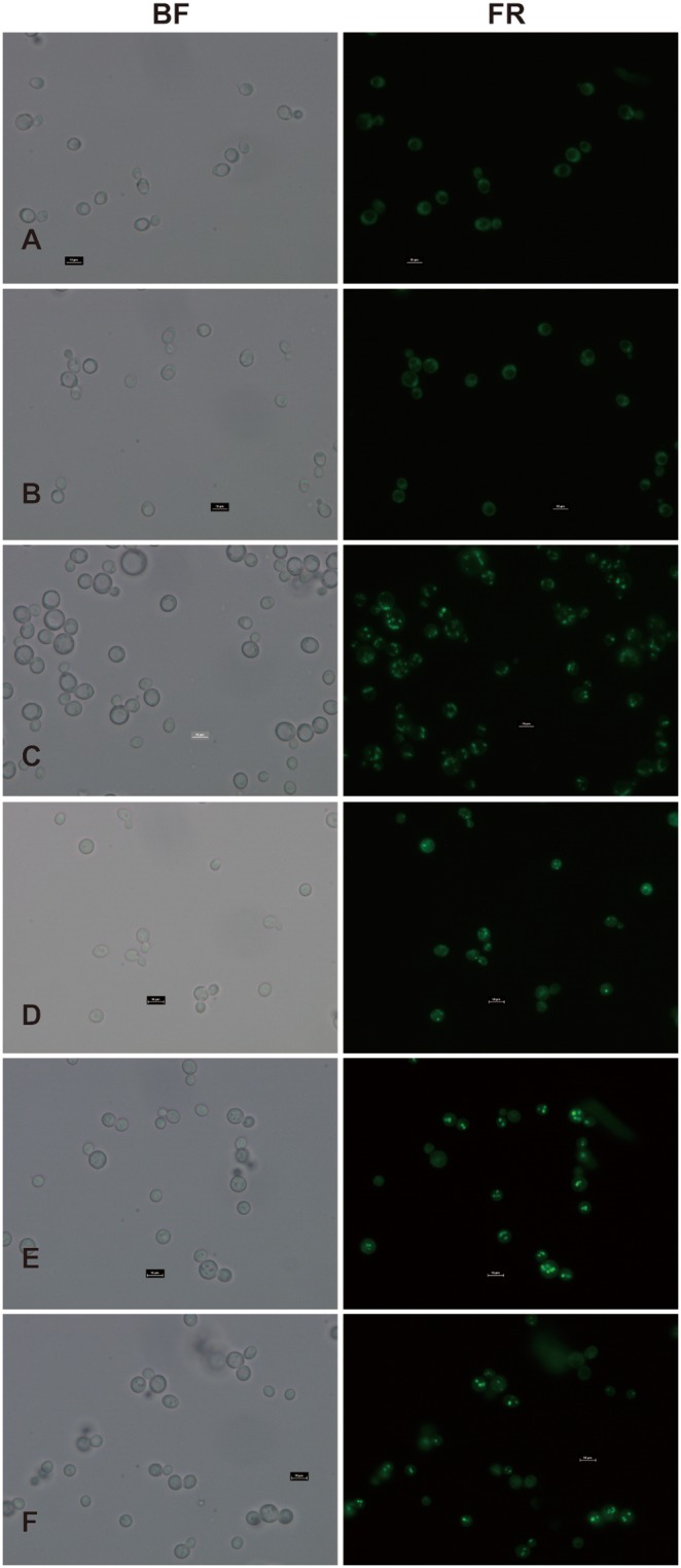
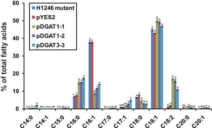
Diacylglycerol acyltransferase (DGAT) catalyzes the final and only committed acylation step in the synthesis of triacylglycerols. In this study, three novel AhDGATs genes were identified and isolated from peanut. Quantitative real-time RT-PCR analysis indicated that the AhDGAT1-2 transcript was more abundant in roots, seeds, and cotyledons, whereas the transcript abundances of AhDGAT1-1 and AhDGAT3-3 were higher in flowers than in the other tissues examined. During seed development, transcript levels of AhDGAT1-1 remained relatively low during the initial developmental stage but increased gradually during later stages, peaking at 50 days after pegging (DAP). Levels of AhDGAT1-2 transcripts were higher at 10 and 60 DAPs and much lower during other stages, whereas AhDGAT3-3 showed higher expression levels at 20 and 50 DAPs. In addition, AhDGAT transcripts were differentially expressed following exposure to abiotic stresses or abscisic acid. The activity of the three AhDGAT genes was confirmed by heterologous expression in a Saccharomyces cerevisiae TAG-deficient quadruple mutant. The recombinant yeasts restored lipid body formation and TAG biosynthesis, and preferentially incorporated unsaturated C18 fatty acids into lipids. The present study provides significant information useful in modifying the oil deposition of peanut through molecular breeding.


Conflict of interest statement

Competing Interests: The authors have declared that no competing interests exist.

Figures

Figure 1
Figure 1. Homology comparison of the amino acid sequences of AhDGAT1 with DGAT1s from other plant species.
Identical amino acid residues are highlighted in black. A putative acyl-CoA binding motif is underlined and designated as block ‘I’. The AS11 tandem repeat is underlined and designated as block ‘II’. The putative catalytic active site is underlined and designated as block ‘III’. The phosphopantetheine attachment site is underlined and designated as block ‘IV’. The SnRK1 target site is designated as block ‘V’. The putative thiolase acyl-enzyme intermediate signature is underlined and designated as block ‘VI’; the dot shows the invariant proline. The putative fatty acid protein signature is underlined and designated as block ‘VII’; the dot shows the tyrosine phosphorylation site. The DAG/phorbol ester binding signature motif is underlined and designated as block ‘VIII’; the dot shows the conserved phenylalanine. The putative C-terminal ER retrieval motifs is underlined and designated as block ‘IX’. The N-glycosylation sites are boxed. Amino acids denoted with triangle represent leucine (L) residues forma putative leucine zipper motif.
Figure 2
Figure 2. Homology comparison of the amino acid sequences of AhDGAT3 with DGAT3s from other plant species.
Identical amino acid residues are highlighted in black. The phosphopantetheine attachment site is underlined and designated as block ‘I’. The potential DGAT motif is underlined and designated as block ‘II’ and ‘V’. The putative thiolase acyl-enzyme intermediate signature is underlined and designated as block ‘III’. The putative Tyr kinase phosphorylation site is underlined and designated as block ‘IV’. The fatty acid binding protein signature is underlined and designated as block ‘VI’. The putative catalytic active site is underlined and designated as block ‘VII’.
Figure 3
Figure 3. Predicted transmembrane domain for peanut DGAT1, DGAT2 and DGAT3 sequences.
The TMHMM web tools (http://www.cbs.dtu.dk/services/TMHMM-2.0/) plot the probability of the ALDH sequence forming a transmembrane helix (0–1.2 on the y-axis). The regions predicted to form transmembrane helix (or helices) are shown in red, while regions of all sequences predicted to be located inside or outside the membrane are shown in blue and pink, respectively. Nine predicted transmembrane helices were identified for AhDGAT1-1 and AhDGAT1-2 sequences, while two transmembrane helix were observed for AhDGAT2 sequences. No transmembrane helice was identified for AhDGAT3-1, AhDGAT3-2 and AhDGAT3-3 sequences.
Figure 4
Figure 4. Phylogenetic tree of DGAT1, DGAT2 and DGAT3 gene families reconstructed by the neighbor-joining (NJ) method.
Gene sequences other than peanut DGATs were shown by their nomenclatures found of www.phytozome.org, with the abbreviations. Colored branches indicated different groups of proteins. Red: DGAT2, blue: DGAT3, purple: DGAT1. Bootstrapping with 1,000 replicates was used to establish the confidence limits of the tree branches.
Figure 5
Figure 5. Expression analysis of three AhDGAT genes using qRT-PCR in seven peanut tissues and at six stages of seed development.
DT (different tissues): R, root; SM, stem; L, leaf; C, cotyledons; H, hypocotyls; F, flower; SD, seed. DS (10 to 60 DAP): six developmental stages of seeds. The relative mRNA abundance was normalized with respect to the peanut AhACT11 gene. The bars were standard deviations (SD) of three technical repeats.
Figure 6
Figure 6. Expression analysis of three AhDGAT genes using qRT-PCR under different stresses.
CL (0 h to 72 h), leaves exposed to cold (4°C) treatment. SL (0 h to 48 h), leaves exposed to high salt (200 mM NaCl) treatment. SR (0 h to 72 h), roots exposed to high salt (200 mM NaCl) treatment. DL (0 h to 72 h), leaves exposed to 20% PEG-6000 treatment. DR (0 h to 72 h), roots exposed to 20% PEG-6000 treatment. AL (0 h to 72 h), leaves exposed to 100 uM ABA treatment. AR (0 h to 72 h), roots exposed to 100 uM ABA treatment. The relative mRNA abundance was normalized with respect to the peanut AhACT11 gene. The bars were standard deviations (SD) of three technical repeats.
Figure 7
Figure 7. Evaluation of TAG biosynthesis in the yeast quadruple mutant (H1246) complemented with AhDGAT genes.
Lipid extracts from the yeast cells were separated by TLC and lipid spots were visualized as described in Materials and Methods. The neutral lipid-deficient quadruple mutant strain H1246 (1) and the mutant harboring the empty vector (pYES2) (2) were used as the negative controls. The wild-type strain INVSc1 was used as a the positive control (3). The quadruple mutant expressing AhDGAT1-1 (4), AhDGAT1-2 (5) and AhDGAT3-3 (6) was analyzed.
Figure 8
Figure 8. Lipid body formation is restored upon expression of AhDGATs in the yeast strain H1246.
Neutral lipid accumulation in lipid bodies was visualized in yeast cells with the fluorescent dye BODIPY505/515. The neutral lipid-deficient quadruple mutant strain H1246 (A) and the mutant harboring the empty vector (pYES2) (B) were used as the negative controls. The wild-type strain INVSc1 was used as a the positive control (C). The quadruple mutant expressing AhDGAT1-1 (D), AhDGAT1-2 (E) and AhDGAT3-3 (F) was analyzed. BF, Bright-field images; FR, images of BODIPY505/515 fluorescence.
Figure 9
Figure 9. Impact of AhDGATs expression on fatty acid profiles of yeast.
The bars were standard deviations (SD) of three technical repeats. For the same fatty acid component, numbers with different letters are statistically significant (P<0.05).

References

    1. Chen X, Snyder CL, Truksa M, Shah S, Weselake RJ (2011) sn-Glycerol-3-phosphate acyltransferases in plants. Plant Signaling and Behavior 6(11): 1695–1699. - PMC - PubMed
    1. Zhang FY, Yang MF, Xu YN (2005) Silencing of DGAT1 in tobacco causes a reduction in seed oil content. Plant Sci 169: 689–694.
    1. Zhang M, Fan J, Taylor DC, Ohlrogge JB (2009) DGAT1 and PDAT1 acyltransferases have overlapping functions in Arabidopsis triacylglycerol biosynthesis and are essential for normal pollen and seed development. Plant Cell 21: 3885–3901. - PMC - PubMed
    1. Ohlrogge J, Browse J (1995) Lipid biosynthesis. Plant Cell 7: 957–970. - PMC - PubMed
    1. Cases S, Novak S, Zheng YW, Myers HM, Lear SR, et al. (1998) ACAT-2, a second mammalian acyl-CoA/cholesterol acyltransferase. Its cloning, expression, and characterization. J Biol Chem 273: 26755–26764. - PubMed

Publication types

MeSH terms

Associated data

LinkOut - more resources