. 2014 Dec;133(12):1497-511.
doi: 10.1007/s00439-014-1481-x. Epub 2014 Sep 3.

Whole exome sequence analysis of Peters anomaly

Affiliations

Whole exome sequence analysis of Peters anomaly

Eric Weh et al. Hum Genet. 2014 Dec.

Abstract

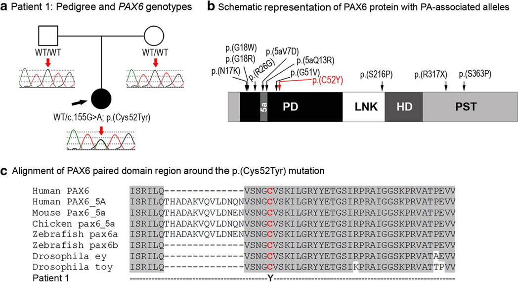
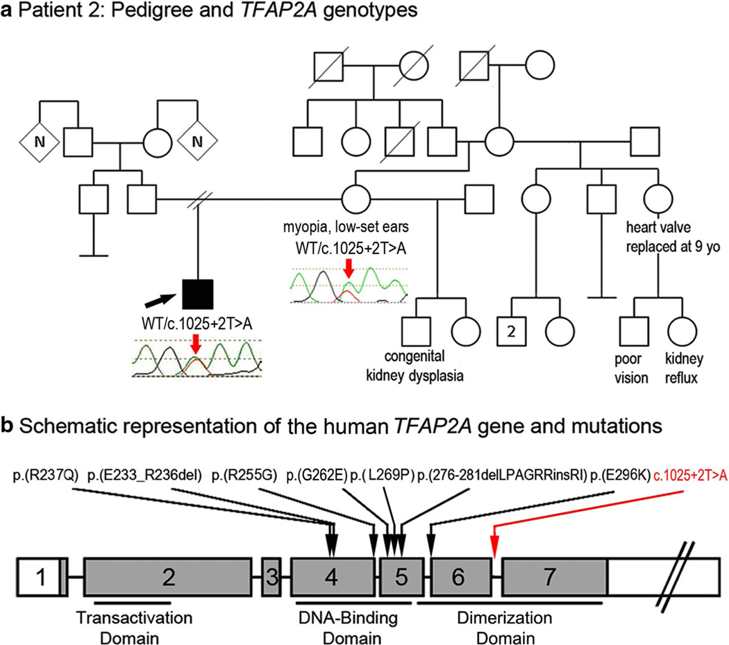
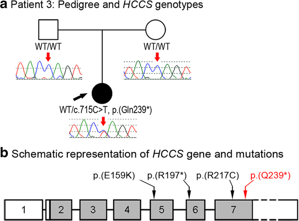
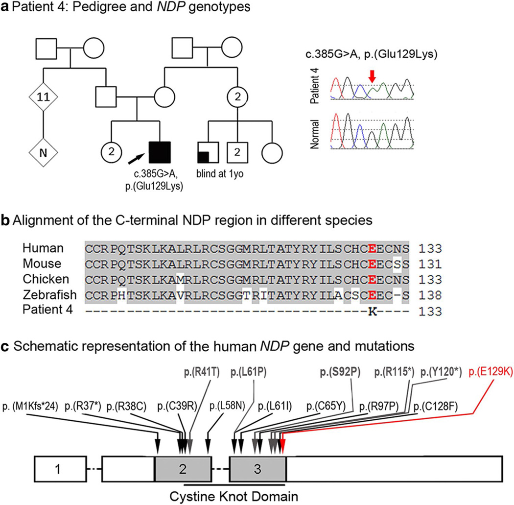
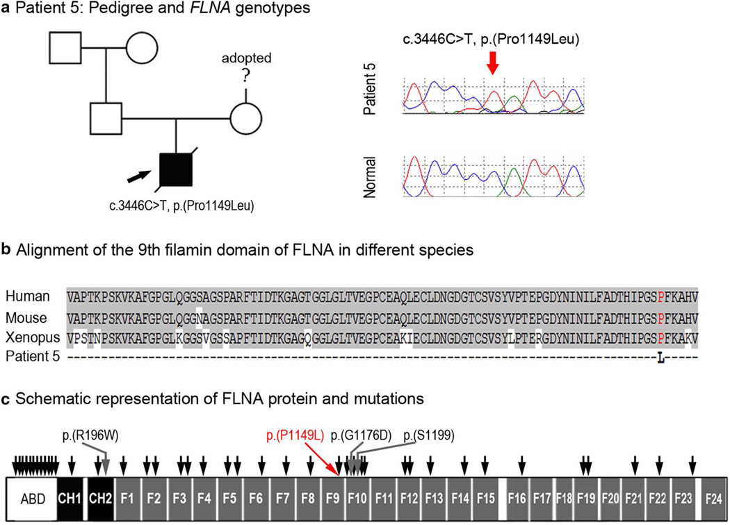
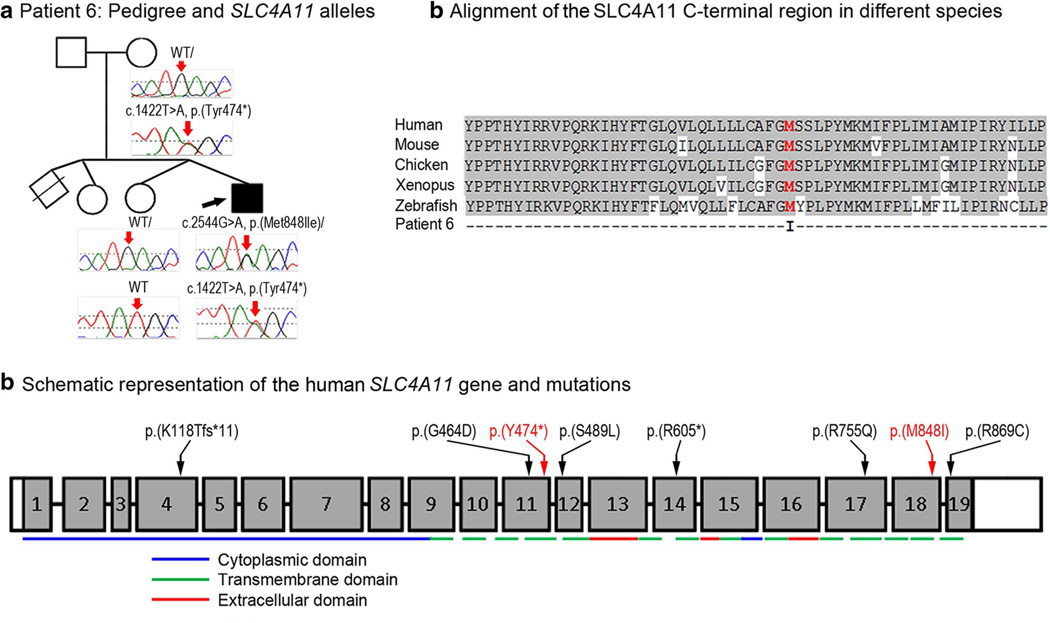
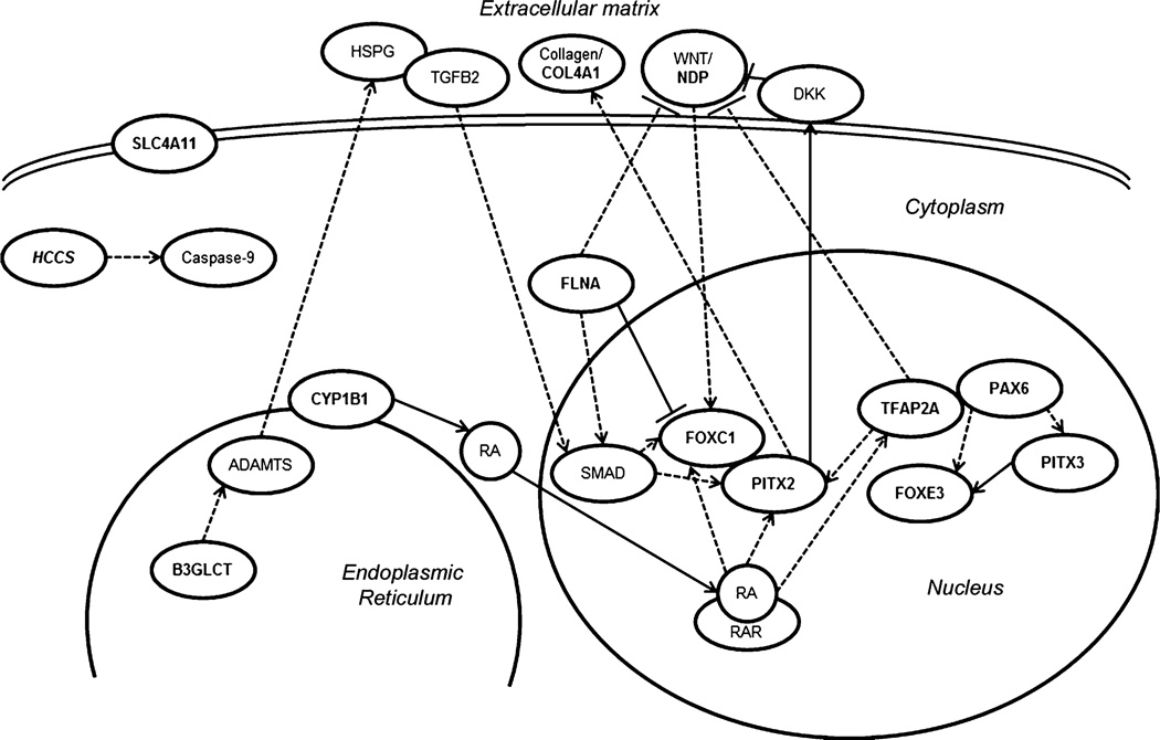
Peters anomaly is a rare form of anterior segment ocular dysgenesis, which can also be associated with additional systemic defects. At this time, the majority of cases of Peters anomaly lack a genetic diagnosis. We performed whole exome sequencing of 27 patients with syndromic or isolated Peters anomaly to search for pathogenic mutations in currently known ocular genes. Among the eight previously recognized Peters anomaly genes, we identified a de novo missense mutation in PAX6, c.155G>A, p.(Cys52Tyr), in one patient. Analysis of 691 additional genes currently associated with a different ocular phenotype identified a heterozygous splicing mutation c.1025+2T>A in TFAP2A, a de novo heterozygous nonsense mutation c.715C>T, p.(Gln239*) in HCCS, a hemizygous mutation c.385G>A, p.(Glu129Lys) in NDP, a hemizygous mutation c.3446C>T, p.(Pro1149Leu) in FLNA, and compound heterozygous mutations c.1422T>A, p.(Tyr474*) and c.2544G>A, p.(Met848Ile) in SLC4A11; all mutations, except for the FLNA and SLC4A11 c.2544G>A alleles, are novel. This is the first study to use whole exome sequencing to discern the genetic etiology of a large cohort of patients with syndromic or isolated Peters anomaly. We report five new genes associated with this condition and suggest screening of TFAP2A and FLNA in patients with Peters anomaly and relevant syndromic features and HCCS, NDP and SLC4A11 in patients with isolated Peters anomaly.


Conflict of interest statement

Conflict of interest The authors declare that they have no relevant financial conflict of interest.

Figures

Fig. 1
Fig. 1
a Patient 1: Pedigree and PAX6 genotypes. DNA chromatograms of the proband’s and parental PAX6 sequences are shown with a position of c.155G>A, p.(Cys52Tyr) mutation indicated with red arrows; proband is designated with a black arrow. b Schematic representation of the PAX6 protein with PA-associated alleles. PD Paired domain, LNK linker region, HD homeodomain, PST transactivation domain rich of proline, serine, and threonine, 5a- 14-a.a. encoded by the alternatively spliced exon 5a. Positions of all previously reported PAX6 mutations associated with Peters anomaly are shown with black arrows; the cysteine residue at position 52 is shown in red font. c Alignment of PAX6 paired domain region around the p.(Cys52Tyr) mutation. Human PAX6 (NP_000271.1), human PAX6-5A (NP_001245391), mouse Pax6-5a (NP_001231127), chicken pax6-5a (NP_990397), zebrafish pax6a (NP_571379), zebrafish pax6b (NP_571716), Drosophila ey (NP_001014693) and Drosophila toy (NP_524638) sequences were used for the alignment; the position of p.(C52Y) is shown with red arrow
Fig. 2
Fig. 2
a Patient 2: Pedigree and TFAP2A genotypes. DNA chromatograms of the proband’s and his mother’s TFAP2A sequences are shown with position of the c.1025+2T>A mutation indicated with red arrows; the proband is designated with a black arrow; phenotypes observed in other members of the pedigree are indicated under corresponding symbols. b Schematic representation of the human TFAP2A gene and mutations. Previously reported mutations (Reiber et al. 2010; Gestri et al. 2009; Tekin et al. 2009) are shown with black arrows; the position of the c.1025+2T>A mutation is shown with a red arrow
Fig. 3
Fig. 3
a Patient 3: pedigree and HCCS genotypes. DNA chromatograms of the proband’s and parental HCCS sequences are shown with the position of the c.715C>T, p.(Gln239*) mutation indicated with red arrows; the proband is designated with a black arrow. b Schematic representation of the human HCCS gene and mutations. HCCS mutations recorded in ClinVar are indicated with black arrows; the position of the p.(Q239*) mutation is shown with a red arrow
Fig. 4
Fig. 4
a Patient 4: pedigree and NDP genotypes. Pedigree is shown on the left, proband is indicated with a black arrow; DNA chromatograms of NDP c.385G>A, p.(Glu129Lys) mutant and normal alleles are shown on the right. b Alignment of the C-terminal NDP region in different species. Human (NP_000257), mouse (NP_035013), chicken (NP_001265015) and zebrafish (UniProt ID: E7F073) sequences were used for the alignment; glutamic acid residue at position 129 is shown in red font. c Schematic representation of the human NDP gene and mutations. Positions of some previously reported NDP mutations associated with Norrie disease or EVR2 are shown with black arrows; positions of mutations previously associated with unspecified corneal opacities are shown at the top and indicated with grey arrows; the p.(E129 K) mutation identified in patient 4 is noted with a red arrow
Fig. 5
Fig. 5
a Patient 5: Pedigree and FLNA genotypes. Pedigree is shown on the left, proband is indicated with a black arrow; DNA chromatograms of FLNA c.3446C>T, p.(Pro1149Leu) mutant and normal alleles are shown on the right. b Alignment of the 9th filamin domain of FLNA in different species. Human (NP_001104026), mouse (NP_001277350), and Xenopus (UniProt ID:F6R7N1) sequences were used for the alignment; proline residue at position 1149 is shown in red font. c Schematic representation of the human FLNA protein and mutations. FLNA domains are indicated according to UniProtKB (P21333): ABD Actin-binding domain, CH1, CH2 calponin homology domains 1 and 2, F1–F24 filamin repeats 1 through 24. The approximate positions of the previously reported FLNA mutations associated with various non-ocular phenotypes are shown by black arrows; mutations associated with ocular phenotypes (please see text) are shown with grey arrows and specified; the p.(P1149L) mutation identified in this study is indicated with red arrow/font
Fig. 6
Fig. 6
a Patient 6: Pedigree and SLC4A11 genotypes. DNA chromatograms of SLC4A11 alleles are shown with positions of c.1422T>A, p.(Tyr474*) and c.2544G>A, p.(Met848Ile) mutations indicated with red arrows; proband is designated with a black arrow. b Alignment of the SLC4A11 C-terminal region in different species. Human (NP_114423), mouse (XP_006499668), chicken (XP_004936340), Xenopus (XM_002936363) and zebrafsh (NM_001159828) sequences were used for the alignment; methionine residue at position 848 is shown in red font. c Schematic representation of the human SLC4A11 gene and mutations. The SLC4A11 exons are shown as numbered grey boxes; positions of cytoplasmic, transmembrane, and extracellular domains encoded by corresponding exonic sequences are indicated at the bottom with blue, green and red lines, correspondingly; positions of the previously described SLC4A11 mutations are indicated with black arrows/font while alleles reported in this study are shown with red arrows/font. SLC4A11 domains and mutations are shown as presented in Vilas et al. (2011) and Vithana et al. (2006)
Fig. 7
Fig. 7
Schematic representation of reported interactions between factors associated with Peters anomaly. The genes implicated in PA based on previous reports as well as this study are marked in bold. PITX2 and FOXC1 have been shown to physically bind and modulate the activity of each other (Acharya et al. 2011) as well as to be regulated by TGFβ (Silla et al. 2014; Iwao et al. 2009), WNT and retinoic acid signaling pathways (Gage and Zacharias 2009; Kumar and Duester 2010; Hatou et al. 2013); in connection with the WNT pathway, PITX2 has been reported to physically bind the DKK2 promoter (Gage and Zacharias 2009). Also, PITX2 has been shown to regulate the deposition of collagens by regulating the procollagen lysyl hydroxylase gene (Hjalt et al. 2001). B3GLCT is a glucosyltransferase which catalyzes the addition of glucose to O-linked fucose via a β-1,3 linkage onto trombospondin type 1 repeats which have been found in the ADAMTS family of proteins (Ricketts et al. 2007; Wang et al. 2009); some ADAMTS proteins have been shown to cleave heparan sulfate proteoglycans (HSPGs) (Barbouri et al. 2014; Kuno and Matsuhima 1998) and thus potentially affect TGFβ signaling (Lin 2004; Iwao et al. 2009). NDP represents a WNT ligand (Ohlmann and Tamm 2012). TFAP2A has been shown to be responsive to retinoic acid, to inhibit the WNT pathway (Li and Dashwood 2004) and be involved in PITX2 regulation (Bamforth et al. 2004). PAX6 deficiency has been shown to negatively affect FOXE3 (Blixt et al. 2007) and PITX3 expression (Chauhan et al. 2002); PITX3 has been reported to directly regulate FOXE3 expression (Shi et al. 2006; Ahmad et al. 2013). PAX6 and TFAP2A have been demonstrated to physically interact and promote transcription of some corneal genes (Sivak et al. 2004). CYP1B1 participates in the synthesis of retinoic acid (Chambers et al. 2007). FLNA appears to negatively regulate FOXC1 by physical binding and sequestering it to heterochromatin and is important for SMAD translocation into the nucleus (Berry et al. 2005; Huang et al. 2009). FLNA may also be important in maintaining appropriate levels of WNT signaling (Adams et al. 2012)

References

    1. Acharya M, Huang L, Fleisch VC, Allison WT, Walter MA. A complex regulatory network of transcription factors critical for ocular development and disease. Hum Mol Genet. 2011;20:1610–1624. - PubMed
    1. Adams M, Simms RJ, Abdelhamed Z, Dawe HR, Szymanska K, Logan CV, Wheway G, Pitt E, Gull K, Knowles MA, Blair E, Cross SH, Sayer JA, Johnson CA. A meckelin-filamin A interaction mediates ciliogenesis. Hum Mol Genet. 2012;21:1272–1286. - PMC - PubMed
    1. Ahmad N, Aslam M, Muenster D, Horsch M, Khan MA, Carlsson P, Beckers J, Graw J. Pitx3 directly regulates Foxe3 during early lens development. Int J Dev Biol. 2013;57:741–751. - PubMed
    1. Bamforth SD, Bragança J, Farthing CR, Schneider JE, Broadbent C, Michell AC, Clarke K, Neubauer S, Norris D, Brown NA, Anderson RH, Bhattacharya S. Cited2 controls left-right patterning and heart development through a Nodal-Pitx2c pathway. Nat Genet. 2004;36:1189–1196. - PubMed
    1. Baratz KH, Tosakulwong N, Ryu E, Brown WL, Branham K, Chen W, Tran KD, Schmid-Kubista KE, Heckenlively JR, Swaroop A, Abecasis G, Bailey KR, Edwards AO. E2-2 protein and Fuchs’s corneal dystrophy. N Engl J Med. 2010;363:1016–1024. - PubMed

Publication types

MeSH terms

Supplementary concepts

LinkOut - more resources