

Anatomical study of minor alterations in neonate vocal folds
- PMID: 25183181
- PMCID: PMC9444660
- DOI: 10.1016/j.bjorl.2014.05.019
Anatomical study of minor alterations in neonate vocal folds
Abstract
Introduction: Minor structural alterations of the vocal fold cover are frequent causes of voice abnormalities. They may be difficult to diagnose, and are expressed in different manners. Cases of intracordal cysts, sulcus vocalis, mucosal bridge, and laryngeal micro-diaphragm form the group of minor structural alterations of the vocal fold cover investigated in the present study. The etiopathogenesis and epidemiology of these alterations are poorly known.
Objective: To evaluate the existence and anatomical characterization of minor structural alterations in the vocal folds of newborns.
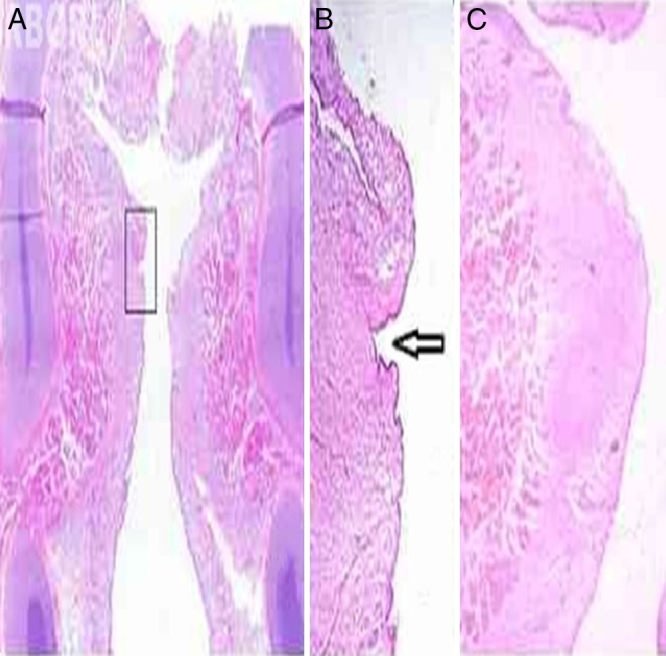
Methods: 56 larynxes excised from neonates of both genders were studied. They were examined fresh, or defrosted after conservation via freezing, under a microscope at magnifications of 25× and 40×. The vocal folds were inspected and palpated by two examiners, with the aim of finding minor structural alterations similar to those described classically, and other undetermined minor structural alterations. Larynges presenting abnormalities were submitted to histological examination.
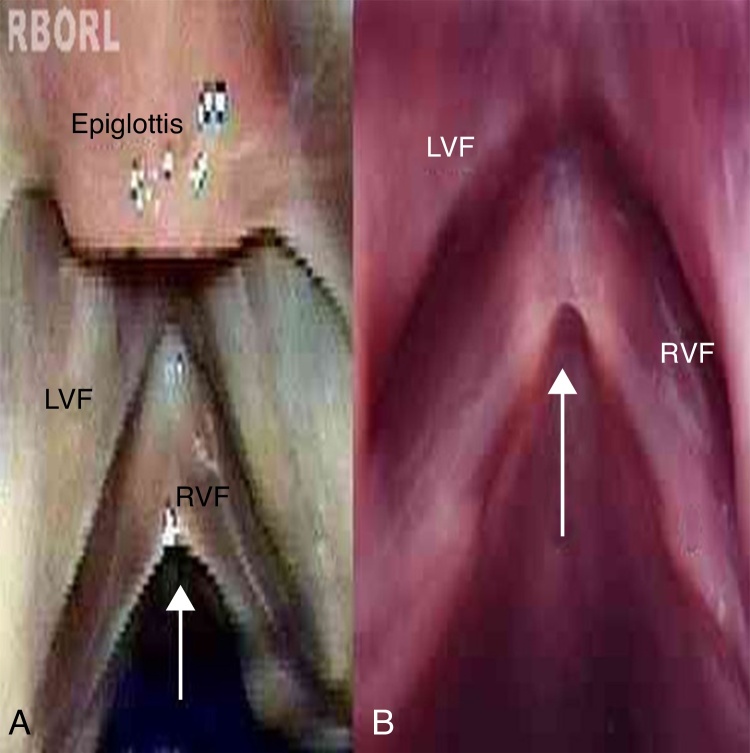
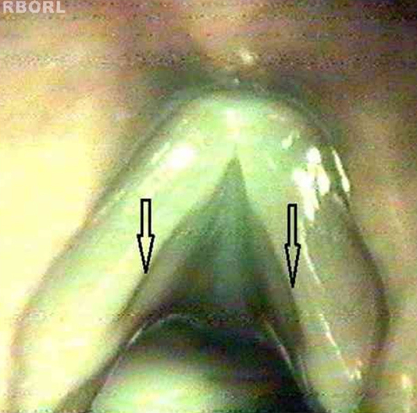
Results: Six cases of abnormalities were found in different larynges: one (1.79%) compatible with a sulcus vocalis and five (8.93%) compatible with a laryngeal micro-diaphragm. No cases of cysts or mucosal bridges were found. The observed abnormalities had characteristics similar to those described in other age groups.
Conclusion: Abnormalities similar to sulcus vocalis or micro-diaphragm may be present at birth.
Introdução: As alterações estruturais mínimas (AEM) da cobertura das pregas vocais são causas frequentes de alterações vocais. Podem ser de diagnóstico difícil, e expressam-se de modo variável. O cisto intracordal, o sulco vocal, a ponte de mucosa e o microdiafragama laríngeo constituem o grupo das AEM da cobertura das pregas vocais pesquisadas neste estudo. Sua etiopatogenia e epidemiologia não são bem conhecidas.
Objetivo: Avaliar a existência e a caracterização anatômica das AEM em prega vocal de neonatos.
Métodos: Foram estudadas 56 laringes excisadas de neonatos, de ambos os sexos. As laringes foram examinadas a fresco ou descongeladas após conservação por congelação, sob microscopia com aumento de 25 e 40×. As pregas vocais foram inspecionadas e palpadas por dois examinadores, com o intuito de encontrar AEM semelhantes às classicamente descritas e outras indeterminadas. As laringes com alterações foram submetidas a exame histológico.
Resultados: Foram encontradas seis alterações em laringes distintas: uma (1,79%) compatível com sulco vocal e cinco (8,93%) compatíveis com microdiafragma laríngeo. Não foram encontrados cistos e pontes de mucosa. As alterações presentes apresentavam características semelhantes às descritas em outras faixas etárias.
Conclusão: Alterações semelhantes ao sulco vocal e ao microdiafragma laríngeo podem estar presentes ao nascimento.
Keywords: Anatomia; Anatomy; Doenças da laringe; Laryngeal diseases; Pregas vocais; Vocal folds.
Copyright © 2014 Associação Brasileira de Otorrinolaringologia e Cirurgia Cérvico-Facial. Published by Elsevier Editora Ltda. All rights reserved.
Figures
References
-
- Arnold G.E. Dysplastic dysphonia: minor anomalies of the vocal cords causing persistent hoarseness. Laryngoscope. 1958;68:142–158. - PubMed
-
- Pontes P., Behlau M. In: Tratado de otorrinolaringologia. Lopes F., Campos C.A.H., editors. Roca; São Paulo: 1994. Disfonias funcionais; pp. 1014–1026.
-
- Pontes P., Behlau M., Gonçalves I. Alterações estruturais mínimas da laringe (AEM): considerações básicas. Acta AWHO. 1994;13:2–6.
-
- Lee S.T.S., Niimi S. Vocal fold sulcus. J Laryngol Otol. 1990;104:876–878. - PubMed
-
- Monday L.A., Cornut G., Bouchayer M., Roch J.B. Epidermoid cysts of the vocal cords. Ann Otol Rhinol Laryngol. 1983;92:124–127. - PubMed
MeSH terms
LinkOut - more resources
Full Text Sources
Other Literature Sources
