Review
. 2015 Jun;9(2):260-8.
doi: 10.1007/s12105-014-0568-y. Epub 2014 Sep 3.

Sarcoidosis of the head and neck

Affiliations
Review

Sarcoidosis of the head and neck

Arvind K Badhey et al. Head Neck Pathol. 2015 Jun.

Abstract

Sarcoidosis is a complex disorder that often times involves the head and neck. Despite the presence of strong clinical evidence, tissue diagnosis and imaging is needed for confirmation of the disease. Although typically managed medically, when found in the sinonasal tract or intracranially, it may necessitate the intervention of a rhinologist-skull base surgeon. This article seeks to provide a comprehensive review of head and neck sarcoidosis, as this fascinating disorder often poses a diagnostic and therapeutic challenge. A brief discussion of surgical treatment for pituitary lesions is also provided. Articles from 1997 to 2013 were selected and reviewed by three researchers utilizing the most recent literature regarding sarcoidosis in the head and neck. PubMed searches were conducted using search terms such as "sarcoidosis", "neurosarcoid", and "extra-pulmonary sarcoid", among many others. A large collection of articles was generated and reviewed by the team of authors, and appropriate information was extracted to compose a thorough and expansive review of the subject. 10-15 % of patients with sarcoidosis have head and neck manifestations. Sinonasal and pituitary sarcoidosis presents a diagnostic challenge owing to its non-specific symptoms. Although systemic steroid therapy is often the first time treatment, endoscopic surgery is commonly used to treat advanced pituitary sarcoidosis refractory to medical management. As tissue diagnosis and imaging is key, a multi-disciplinary team approach is advantageous. Our study collates the available literature on head and neck sarcoidosis to provide a comprehensive review of the subject. This provides helpful information to guide all practitioners involved in the care of these challenging patients, namely pathologists, radiologists, otolaryngologists, and skull base surgeons, in the workup and management of head and neck sarcoidosis.
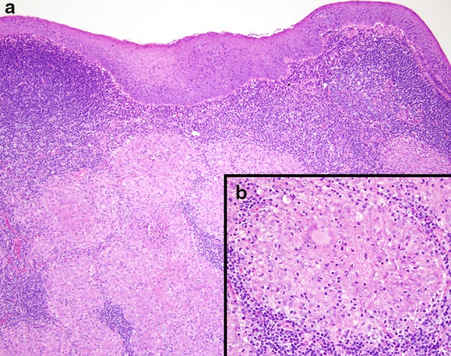

Figures

Fig. 1
Fig. 1
a 10 × magnification of a nasopharyngeal specimen in a patient with sarcoidosis showing a well formed granuloma with lymphoid hyperplasia in an inflammatory background (Original image provided courtesy of Dr. Codrin Iacob, Department of Pathology at New York Eye and Ear Infirmary-Mount Sinai Health System). b 40 × magnification of a granuloma isolated from a nasopharyngeal specimen. Note the presence of a well developed giant cell histiocyte. (Original image provided courtesy of Dr. Codrin Iacob, Department of Pathology at New York Eye and Ear Infirmary-Mount Sinai Health System)
Fig. 2
Fig. 2
20 × magnification of laryngeal specimen showing granuloma in a patient with sarcoidosis (Original image provided courtesy of Dr. Codrin Iacob, Department of Pathology at New York Eye and Ear Infirmary-Mount Sinai Health System)
Fig. 3
Fig. 3
40 × magnification of the same laryngeal specimen seen previously in Fig. 7 (Original image provided courtesy of Dr. Codrin Iacob, Department of Pathology at New York Eye and Ear Infirmary-Mount Sinai Health System)
Fig. 4
Fig. 4
Post contrast coronal T1 weighted image showing bilateral temporal leptomeningeal enhancement, enhancement of the optic chiasm, and bilateral cavernous sinus enhancement. (Original image provided courtesy of Dr. Azita Khorsandi, Department of Radiology at New York Eye and Ear Infirmary-Mount Sinai Medical Center)
Fig. 5
Fig. 5
Post contrast sagittal T1 weighted image demonstrating suprasellar cistern enhancement along the optic chiasm and radiation. Frontal leptomeningeal enhancement is also noted (Original image provided courtesy of Dr. Azita Khorsandi, Department of Radiology at New York Eye and Ear Infirmary-Mount Sinai Medical Center)
Fig. 6
Fig. 6
20 × image of a lacrimal gland specimen showing mucinous and serious acini, along with granuloma with chronic inflammation (Original image provided courtesy of Dr. Codrin Iacob, Department of Pathology at New York Eye and Ear Infirmary-Mount Sinai Health System)
Fig. 7
Fig. 7
Post contrast coronal T1 weighted image demonstrating cerebellar and frontal nodular leptomeningeal enhancement (Original image provided courtesy of Dr. Azita Khorsandi, Department of Radiology at New York Eye and Ear Infirmary-Mount Sinai Medical Center)

References

    1. Dash GI, Kimmelman CP. Head and neck manifestations of sarcoidosis. The Laryngoscope. 1988;98(1):50–53. doi: 10.1288/00005537-198801000-00011. - DOI - PubMed
    1. Shah UK, White JA, Gooey JE, Hybels RL. Otolaryngologic manifestations of sarcoidosis: presentation and diagnosis. The Laryngoscope. 1997;107(1):67–75. doi: 10.1097/00005537-199701000-00015. - DOI - PubMed
    1. Schwartzbauer HR, Tami TA. Ear, nose, and throat manifestations of sarcoidosis. Otolaryngol clin N Am. 2003;36(4):673–684. doi: 10.1016/S0030-6665(03)00030-6. - DOI - PubMed
    1. Mrowka-Kata K, Kata D, Lange D, Namyslowski G, Czecior E, Banert K. Sarcoidosis and its otolaryngological implications. Eur Arch Otorhinolaryngol. 2010;267(10):1507–1514. doi: 10.1007/s00405-010-1331-y. - DOI - PubMed
    1. Dastoori M, Fedele S, Leao JC, Porter SR. Sarcoidosis: a clinically orientated review. J Oral Pathol Med. 2013;42(4):281–289. doi: 10.1111/j.1600-0714.2012.01198.x. - DOI - PubMed

MeSH terms

Supplementary concepts