. 2014;42(17):11166-79.
doi: 10.1093/nar/gku758. Epub 2014 Sep 2.

Processing of the seven valine tRNAs in Escherichia coli involves novel features of RNase P

Affiliations

Processing of the seven valine tRNAs in Escherichia coli involves novel features of RNase P

Ankit Agrawal et al. Nucleic Acids Res. 2014.

Abstract

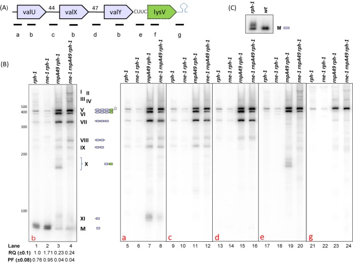
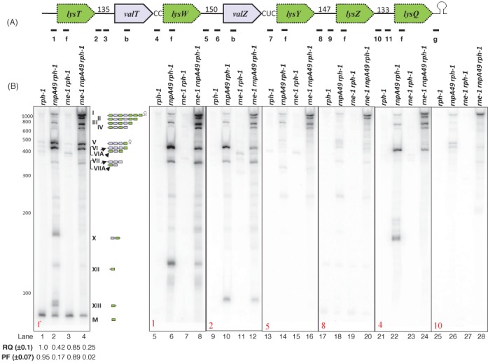
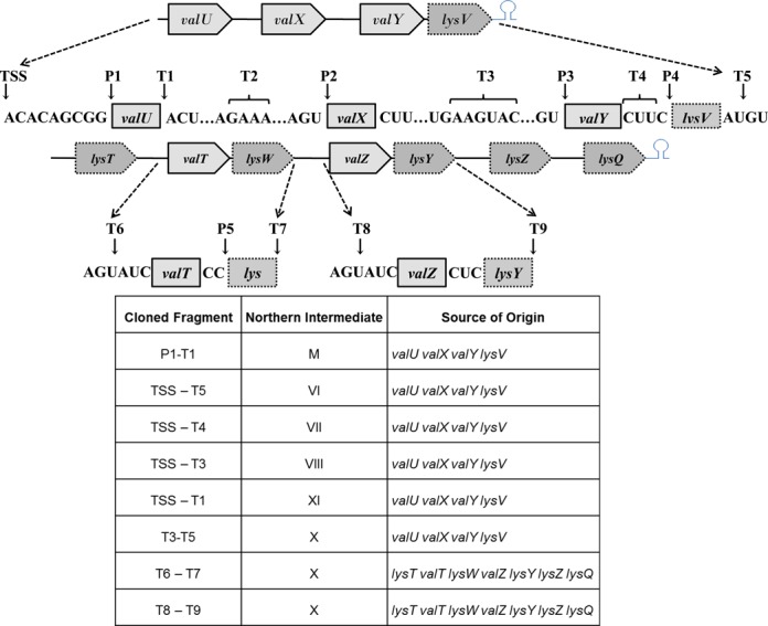
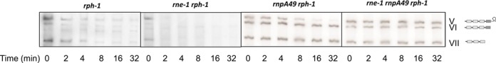
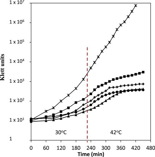
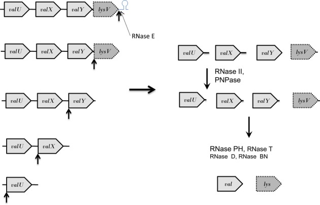
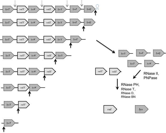
Here we report that RNase P is required for the initial separation of all seven valine tRNAs from three distinct polycistronic transcripts (valV valW, valU valX valY lysY and lysT valT lysW valZ lysY lysZ lysQ). Particularly significant is the mechanism by which RNase P processes the valU and lysT polycistronic transcripts. Specifically, the enzyme initiates processing by first removing the Rho-independent transcription terminators from the primary valU and lysT transcripts. Subsequently, it proceeds in the 3' → 5' direction generating one pre-tRNA at a time. Based on the absolute requirement for RNase P processing of all three primary transcripts, inactivation of the enzyme leads to a > 4-fold decrease in the levels of both type I and type II valine tRNAs. The ability of RNase P to initiate tRNA processing at the 3' ends of long primary transcripts by endonucleolytically removing the Rho-independent transcription terminator represents a previously unidentified function for the enzyme, which is responsible for generating the mature 5' termini of all 86 E. coli tRNAs. RNase E only plays a very minor role in the processing of all three valine polycistronic transcripts.


Figures

Figure 1.
Figure 1.
Analysis of valU operon processing. (A) Schematic diagram of the valU operon (not drawn to scale). Relative positions of the oligonucleotide probes (a: valU-UP, b: valUXYTZ, c: valU-X, d: valX-Y, e: valY-lysV, f: lysmature, g:lysV-TER) used in Northern analyses are shown. Probe sequences are listed in Supplementary Table S1 (Supplementary Materials). Numbers indicate the length of the intergenic spacers. (B) Northern analysis of valU operon. The specific oligonucleotide probe used for each blot is indicated in the bottom left corner of each blot. The genotypes of the strains used are listed on the top of each autoradiogram. The RNA molecular size standards (nucleotides) (Fermentas) are shown to the left of the first image. The numerical designations of the different species along with their graphical structures are shown to the right of the first blot. Relative quantity (RQ) of mature tRNAVal (M) in various genetic backgrounds was calculated by setting a level of one in the rph-1 strain. Processed fraction (PF) represents the fraction of mature tRNA relative to the total amount of processed and unprocessed species of that tRNA. Each value represents the average of at least three independent determinations. (C) Analysis of the role of RNase PH in the final maturation of the five valine type I tRNAs.
Figure 2.
Figure 2.
Analysis of the lysT operon processing. (A) Schematic diagram of the operon (not drawn to scale). Relative positions of the oligonucleotide probes (1: lysT-UP, f: lysmature, 2: lysT-valT1, 3: lysT-valT3, b: valUXYTZ, 4: valT-lysW, 5: lysW-valZ1, 6: valZ-UP2, 7: valZ-lysY, 8: lysY-lysZ1, 9: lysY-lysZ2, 10: lysZ-lysQ1, 11: lysZ-lysQ2, g: lysV-TER). Probe sequences are listed in Supplementary Table S1 (Supplementary Materials). Numbers indicate the length of the intergenic spacers. (B) Northern analysis of lysT operon. The specific oligonucleotide probe used in each blot is indicated in the bottom left corner of the blot. The genotypes of the strains used are indicated on the top of the each autoradiogram. The RNA molecular size standards (nucleotides) (Fermentas) are shown to the left of the first blot. The numerical designations of the different species along with their graphical structures are shown to the right of the blot. Bands VI and VII arise from the valU operon and comigrate with bands VIA and VIIA, respectively. Two independent Northern blots were run (one was probed with f, 1, 2, 5 and 8; second was probed with 4 and 10). The RQ and PF values were calculated as described in the legend to Figure 1.
Figure 3.
Figure 3.
Identification of cleavage sites within the valU and lysT operons by cloning and sequencing of processing intermediates. cDNAs of self-ligated RNA molecules were amplified, cloned and sequenced to identify the 5′ and 3′ ends as described in ‘Materials and Methods’. Each arrow represents either a 5′ or 3′ end based on the DNA sequence. Multiple ends were obtained at the T2, T3 and T4 locations. The transcription, start-site (TSS), RNase P cleavage sites (P1–5), 3′ cleavage sites [either at the mature 3’ end or in the intergenic region for respective upstream tRNAs (T1–5, 7 and 9)] and 5′ cleavage sites (in the intergenic regions) for respective downstream tRNAS (T6, 8) are indicated. The 3′ and 5′ ends of some of the major processing intermediates observed in the Northern analyses (Figures 1 and 2) are shown below the diagram.
Figure 4.
Figure 4.
Northern analysis of the decay of valU operon. The genotypes of the strains are indicated on the top and time-points are shown at the bottom of the autoradiograms. The blots were probed with probe b (Figure 1A). The numerical designations of the different species with their graphical structures are shown to the right and are identical to those shown in Figure 1.
Figure 5.
Figure 5.
Growth curve of various strains. All strains were initially grown at 30°C until 50 Klett units above background when IPTG was added to a final concentration of 350 μM. The cultures were diluted with pre-warmed Luria broth (LB)/thymine or LB/thymine/IPTG to maintain them in exponential growth (Klett 50–70). For this experiment, all the cultures were shifted to 42°C when the slowest growing culture reached Klett 50 above background (vertical dashed line), since it has been previously shown that overproduction of rnpB does not complement at 44°C (36). The optical densities (OD) were measured with a Klett-Summerson Colorimeter (No. 42 Green filter). x: MG1693 (rph-1); ♦: SK2525 (rnpA49 rph-1); ▴: SK10537 (rnpA49 rph-1 argX+); •: SK10541 (rnpA49 rph-1 valU+valW+); ▪: SK10539 (rnpA49 rph-1 rnpB+).
Figure 6.
Figure 6.
Processing pathway for the valU polycistronic transcript. Similar to what has been observed with the valV valW and secG leuU primary transcripts (15,17), RNase P (upward black arrows) is the primary processing enzyme for the valU transcript. Initially, RNase P efficiently removes the Rho-independent transcription terminator (upward black arrow). Subsequent sequential cleavages occur in the 3′ → 5′ direction at the mature 5′ termini of lysV, valY, valX and valU (upward black arrows). The valU and valX pre-tRNAs will have long 3′ termini of 44 and 47 nt, respectively (Figure 1A) that will be initially processed by a combination of RNase II and PNPase (17,48). Subsequently, the final processing of the valine pre-tRNAs will be carried out primarily by RNase PH because of the presence of C residues downstream of the CCA determinant (42). The final processing of the lysV pre-tRNA employs RNase T. In the absence of RNase P, RNase E can inefficiently remove (dashed arrow) the Rho-independent transcription terminator (Figure 1B). Transcripts are not drawn to scale.
Figure 7.
Figure 7.
Processing pathway for the lysT polycistronic operon. Similar to what was described in Figure 6, after RNase P removes the Rho-independent transcription terminator (upward black arrow), subsequent sequential cleavages will occur in the 3′ → 5′ direction at the mature 5′ termini of lysQ, lysZ, lysZ, lysW, valT and lysT (upward black arrows) to generate pre-tRNAs containing between 2 and150 nt extra nucleotides at their 3′ ends (Figure 2A). The long 3′ ends of the five lys pre-tRNAs will be initially processed by a combination of RNase II and PNPase (17,48). Final maturation of the lys tRNAs primarily is carried out by RNase T, while the final processing of the two valine species is done by RNase PH. In the absence of RNase P, RNase E (dashed downward arrows) can cleave within the long spacer regions between lysT and valT, lysW and valZ, lysY and lysZ, and lysZ and lysQ. However, RNase E does not separate valT and lysW or valZ and lysY. Transcripts are not drawn to scale.

References

    1. Blattner F.R., Plunkett G., III, Bloch C.A., Perna N.T., Burland V., Riley M., Collado-Vides J., Glasner J.D., Rode C.K., Mayhew G.F., et al. The complete sequence of Escherichia coli K-12. Science. 1997;277:1453–1474. - PubMed
    1. Berlyn M.K.B. Linkage map of Escherichia coli K-12, edition 10: the traditional map. Microbiol. Mol. Biol. Rev. 1998;62:814–984. - PMC - PubMed
    1. Li Z., Deutscher M.P. RNase E plays an essential role in the maturation of Escherichia coli tRNA precursors. RNA. 2002;8:97–109. - PMC - PubMed
    1. Ow M.C., Kushner S.R. Initiation of tRNA maturation by RNase E is essential for cell viability in E. coli. Genes Dev. 2002;16:1102–1115. - PMC - PubMed
    1. Deutscher M.P. Degradation of RNA in bacteria: comparison of mRNA and stable RNA. Nucleic Acids Res. 2006;34:659–666. - PMC - PubMed

Publication types

MeSH terms

LinkOut - more resources