

Advanced magnetic resonance imaging of the physical processes in human glioblastoma
- PMID: 25183787
- PMCID: PMC4155518
- DOI: 10.1158/0008-5472.CAN-14-0383
Advanced magnetic resonance imaging of the physical processes in human glioblastoma
Abstract
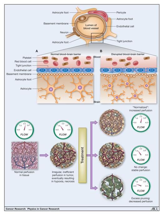
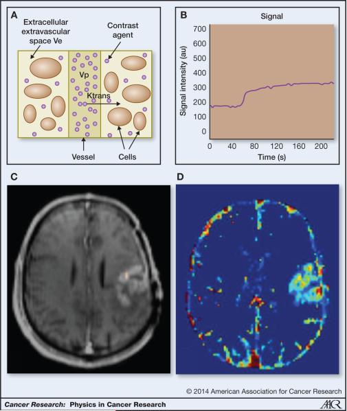
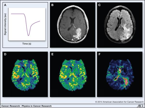
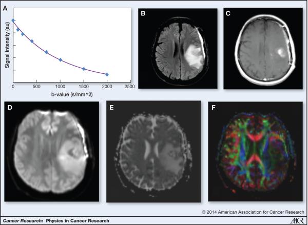
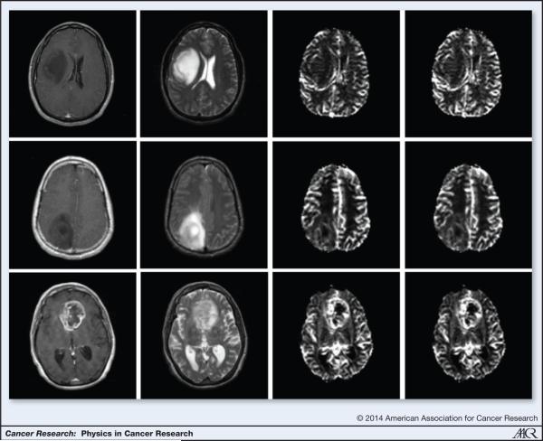
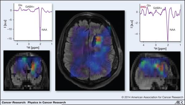
The most common malignant primary brain tumor, glioblastoma multiforme (GBM) is a devastating disease with a grim prognosis. Patient survival is typically less than two years and fewer than 10% of patients survive more than five years. Magnetic resonance imaging (MRI) can have great utility in the diagnosis, grading, and management of patients with GBM as many of the physical manifestations of the pathologic processes in GBM can be visualized and quantified using MRI. Newer MRI techniques such as dynamic contrast enhanced and dynamic susceptibility contrast MRI provide functional information about the tumor hemodynamic status. Diffusion MRI can shed light on tumor cellularity and the disruption of white matter tracts in the proximity of tumors. MR spectroscopy can be used to study new tumor tissue markers such as IDH mutations. MRI is helping to noninvasively explore the link between the molecular basis of gliomas and the imaging characteristics of their physical processes. We, here, review several approaches to MR-based imaging and discuss the potential for these techniques to quantify the physical processes in glioblastoma, including tumor cellularity and vascularity, metabolite expression, and patterns of tumor growth and recurrence. We conclude with challenges and opportunities for further research in applying physical principles to better understand the biologic process in this deadly disease. See all articles in this Cancer Research section, "Physics in Cancer Research."
©2014 American Association for Cancer Research.
Figures
Similar articles
-
Imaging biomarkers guided anti-angiogenic therapy for malignant gliomas.Neuroimage Clin. 2018 Jul 5;20:51-60. doi: 10.1016/j.nicl.2018.07.001. eCollection 2018. Neuroimage Clin. 2018. PMID: 30069427 Free PMC article. Review.
-
Glioblastoma Exhibits Inter-Individual Heterogeneity of TSPO and LAT1 Expression in Neoplastic and Parenchymal Cells.Int J Mol Sci. 2020 Jan 17;21(2):612. doi: 10.3390/ijms21020612. Int J Mol Sci. 2020. PMID: 31963507 Free PMC article.
-
Combination of IVIM-DWI and 3D-ASL for differentiating true progression from pseudoprogression of Glioblastoma multiforme after concurrent chemoradiotherapy: study protocol of a prospective diagnostic trial.BMC Med Imaging. 2017 Feb 1;17(1):10. doi: 10.1186/s12880-017-0183-y. BMC Med Imaging. 2017. PMID: 28143434 Free PMC article. Clinical Trial.
-
Radiotherapy of high-grade gliomas: current standards and new concepts, innovations in imaging and radiotherapy, and new therapeutic approaches.Chin J Cancer. 2014 Jan;33(1):16-24. doi: 10.5732/cjc.013.10217. Chin J Cancer. 2014. PMID: 24384237 Free PMC article. Review.
-
Advanced magnetic resonance imaging in glioblastoma: a review.Chin Clin Oncol. 2017 Aug;6(4):40. doi: 10.21037/cco.2017.06.28. Chin Clin Oncol. 2017. PMID: 28841802 Review.
Cited by
-
Diffusion Weighted Imaging in Neuro-Oncology: Diagnosis, Post-Treatment Changes, and Advanced Sequences-An Updated Review.Cancers (Basel). 2023 Jan 19;15(3):618. doi: 10.3390/cancers15030618. Cancers (Basel). 2023. PMID: 36765575 Free PMC article. Review.
-
The Role of Advanced Brain Tumor Imaging in the Care of Patients with Central Nervous System Malignancies.Curr Treat Options Oncol. 2018 Jun 21;19(8):40. doi: 10.1007/s11864-018-0558-5. Curr Treat Options Oncol. 2018. PMID: 29931476 Review.
-
Molecular Markers of Therapy-Resistant Glioblastoma and Potential Strategy to Combat Resistance.Int J Mol Sci. 2018 Jun 14;19(6):1765. doi: 10.3390/ijms19061765. Int J Mol Sci. 2018. PMID: 29899215 Free PMC article. Review.
-
Radioiodinated PARP1 tracers for glioblastoma imaging.EJNMMI Res. 2015 Dec;5(1):123. doi: 10.1186/s13550-015-0123-1. Epub 2015 Sep 4. EJNMMI Res. 2015. PMID: 26337803 Free PMC article.
-
Multimodal MRI characteristics of the glioblastoma infiltration beyond contrast enhancement.Ther Adv Neurol Disord. 2019 May 14;12:1756286419844664. doi: 10.1177/1756286419844664. eCollection 2019. Ther Adv Neurol Disord. 2019. PMID: 31205490 Free PMC article.
References
-
- Stupp R, Hegi ME, Mason WP, van den Bent MJ, Taphoorn MJ, Janzer RC, et al. Effects of radiotherapy with concomitant and adjuvant temozolomide versus radiotherapy alone on survival in glioblastoma in a randomised phase III study: 5-year analysis of the EORTC-NCIC trial. The lancet oncology. 2009;10:459–66. - PubMed
-
- Wen PY, Kesari S. Malignant gliomas in adults. N Engl J Med. 2008;359:492–507. - PubMed
Publication types
MeSH terms
Substances
Grants and funding
LinkOut - more resources
Full Text Sources
Other Literature Sources
Medical
