Review
. 2014 Sep 2;6(9):a016006.
doi: 10.1101/cshperspect.a016006.

The neomuran revolution and phagotrophic origin of eukaryotes and cilia in the light of intracellular coevolution and a revised tree of life

Affiliations
Review

The neomuran revolution and phagotrophic origin of eukaryotes and cilia in the light of intracellular coevolution and a revised tree of life

Thomas Cavalier-Smith. Cold Spring Harb Perspect Biol. .

Abstract

Three kinds of cells exist with increasingly complex membrane-protein targeting: Unibacteria (Archaebacteria, Posibacteria) with one cytoplasmic membrane (CM); Negibacteria with a two-membrane envelope (inner CM; outer membrane [OM]); eukaryotes with a plasma membrane and topologically distinct endomembranes and peroxisomes. I combine evidence from multigene trees, palaeontology, and cell biology to show that eukaryotes and archaebacteria are sisters, forming the clade neomura that evolved ~1.2 Gy ago from a posibacterium, whose DNA segregation and cell division were destabilized by murein wall loss and rescued by the evolving novel neomuran endoskeleton, histones, cytokinesis, and glycoproteins. Phagotrophy then induced coevolving serial major changes making eukaryote cells, culminating in two dissimilar cilia via a novel gliding-fishing-swimming scenario. I transfer Chloroflexi to Posibacteria, root the universal tree between them and Heliobacteria, and argue that Negibacteria are a clade whose OM, evolving in a green posibacterium, was never lost.


Figures

Figure 1.
Figure 1.
Relationships between the five major cell types, showing key evolutionary innovations in the transitions making them. Rigid murein cell walls originated before the cenancestor of all life using both d- and l-amino acids in the first cell, a posibacterium with acyl ester glycerophospholipids that divided using FtsZ, possibly a photoheterotroph similar to Heliobacterium. Negibacteria evolved by acquiring an outer membrane (OM) with complex targeting of porins and other β-barrel proteins inserted by Omp85-dependent machinery never lost in the history of life, being retained when eukaryotes enslaved phagocytosed negibacteria to make mitochondria and subsequently chloroplasts (even kept in secondarily anaerobic DNA-free hydrogenosomes and mitosomes that evolved by drastically modifying aerobic mitochondria). The neomuran revolution was arguably a stabilizing response to traumatic loss of murein. Histones H3/4 ensured passive negative DNA supercoiling (making nucleosomes) to replace eubacterial ATP-driven supercoiling by DNA gyrase; this stabler DNA coiling forced drastic coevolutionary changes in RNA polymerase and especially DNA replication machinery: repair polymerase δ replaced DNA polymerase III, the β-clamp became PCNA, the replication fork helicase Mcm replaced DnaB, the unrelated Pol primase replaced DnaG primase, and Cdc6 replaced the replication initiator DnaA; Cdc6 possibly evolved from a gene duplicate of the eubacterial clamp loader DnaX, itself undergoing minor modification to neomuran RFC. Novel TATA-box-binding transcription factors (TBP and others) (Ouhammouch et al. 2009) replaced the eubacterial transcription regulator CrtA. Murein loss freed MreB filaments that maintain eubacterial rod shape (or related ParM filaments that segregate some plasmids) to become the actin endoskeleton, conferring osmotic stability; new ESCRT-III filaments helped membrane division, allowing loss of FtsZ in eukaryotes and some archaebacteria. Novel cotranslationally made N-linked glycoprotein enabled archaebacteria to make rigid S-layer-like walls and eukaryotes a flexible cell surface coat, allowing phagotrophy and ingestion of prey cells to evolve, triggering a cascade of eukaryogenic changes associated with coated vesicle origins. These mediated endomembrane differentiation, internal digestion, targeted vesicle fusion, and nuclear envelope evolution to protect chromatin internalized by phagocytosis (see Fig. 2); α-tubulin, β-tubulin, and γ-tubulin evolved from posibacterial plasmid-segregating TubZ GTPase, enabling DNA segregation by mitosis, drastically changing chromosome organization; cohesins enabling mitosis and eukaryotic cell-cycle controls evolved from duplicated Smc condensins. Archaebacteria replaced acyl ester lipids by heat-stable isoprenoid tetraethers to become the first extremophiles, but lost so many lipids and proteins that they could never have evolved directly into eukaryotes, as did the transient neomuran ancestor, retaining far more eubacterial characters. Archaebacteria kept fatty acid (FA) synthesis (Lombard et al. 2012a) but lost acyl-carrier protein (ACP), which enables rapid bulk FA synthesis in eubacteria and eukaryotes, no longer needed for the trace FA amounts that sufficed after archaebacteria lost acyl esters, including phosphatidylinositol (PI) and cardiolipin (CL). Neokaryotes retained bacterial transcription regulation but evolved new transcription factors (TFs). Soon after mitochondrially donated group II self-splicing introns became spliceosomal introns in the cenancestral eukaryote (Cavalier-Smith 1991c), Euglenozoa evolved trans-splicing of spliced-leader (SL) miniexons for all mRNAs (Cavalier-Smith 1993) and lost transcriptional control of gene expression. Neokaryotes alone replaced centromeric histone H3 by CENP-A and evolved Smc5/6 for DNA repair. Archaebacterial flagella are not homologous to, and evolved independently of, eubacterial flagella, which must have evolved in Negibacteria, probably in early Gracilicutes (Cavalier-Smith 2006c); so if the tree is correctly rooted within Posibacteria, they were presumably acquired by Posibacteria by lateral gene transfer (LGT) subsequently, but before actinobacteria diverged from Teichobacteria (see Fig. 3). Ancestral green bacteria lacked flagella but could probably glide and thus make stromatolites, yielding the oldest fossil evidence for eubacteria. Absence of photosynthetic carbon fixation in archaebacteria means that, unlike the much older eubacteria, they could never have fueled an extensive global ecosystem alone.
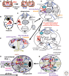
Figure 2.
Figure 2.
Intracellular coevolution during phagotrophy-driven eukaryogenesis. (A) Eubacteria segregate DNA by actively moving replicon origins (O) by ParABS machinery, DNA condensation by Smc condensin rings, and moving termini (T) by the DNA translocase FtsK anchored at the mid-cell nascent division site, marked by the GTPase FtsZ ring for membrane scission by the divisome after XerCD recombinase resolves daughter DNAs into covalently separate molecules. Neomuran loss of murein disrupted orderly linear arrangement of chromosome origins and termini on a rigid wall, causing FtsK loss and allowing replicon numbers per chromosome to increase, and ESCRT-III GTPase filaments replaced the eubacterial divisome; simultaneously, the posibacterial paracrystalline S layer became novel N-linked glycoproteins. (B) In eukaryotes only, these glycoproteins (yellow) became flexible and specialized for binding prey, initially digested by enzymes secreted externally by membrane-attached ribosomes. MreB (or its plasmid segregation ParM relative) (Yutin et al. 2009) evolved into actin, yielding linear filaments stimulated by formins for extending cytoplasm partly around prey, thereby increasing digestion product absorption, and duplications yielded Arp2/3, generating an osmotically stabilizing branching endoskeleton (blue). (C) Evolution of a surface membrane protein channel (Derlin) enabled partially digested proteins to be pulled across the membrane and fully digested on its cytosolic face by cylindrical proteasomes. Digestion products of prey completely internalized into a phagosome (center) was most efficiently absorbed; phagosome-associated V-SNAREs and CM-associated T-SNAREs evolved to refuse phagosome membranes with the surface. (D) Accidental phagocytic internalization of membrane-attached DNA was made permanent by evolving COP-coated vesicles that returned membrane only to the cell surface; after exclusion of ribosomes and DNA from COP vesicles, continued phagocytosis removed all from the plasma membrane, and the internalized ribosome/DNA-associated membrane became protoER. Membrane fragmentations generated separate compartments specializing in β-oxidation of fatty acids (peroxisomes) and cytochrome P450 oxidation of aromatics and protein secretion (ER). Bacterial Sec61/SRP for extruding unfolded proteins was retained by ER and TAT machinery for unfolded proteins modified for peroxisome biogenesis (for more details, see Cavalier-Smith 2009). FtsZ-related posibacterial plasmid-segregated TubZ GTPase evolved by gene duplication into eukaryotic mitotic segregator (α-,β-tubulin microtubules, nucleated at minus ends by γ-tubulin-containing centrosomes), microtubule rigidity mechanistically replacing peptidoglycan rigidity. Initially, microtubule polymerization forced sister centrosomes and associated DNA apart. COPs were also used for pinocytosis, and preexisting dynamin and ESCRT-III coopted for membrane scission generating protoendosomes (pE). Single-head myosin I and kinesin diverged from a common posibacterial ATPase ancestor to form motors for internalizing phagosomes along actin filaments or minus-to-plus movement of vesicles on microtubules for exocytosis, respectively. (E) Kinesin was coopted to push apart antiparallel microtubules from sister centrosomes, improving segregation, cytokinesis being by preexisting neomuran ESCRT-III GTPase ring orthogonal to the spindle. (F) Simultaneously, coordinate gene duplications of COP proteins and SNAREs multiplied the number of topologically and chemically distinct compartments developmentally interlinked by vesicle transport: copII for ER to Golgi, CopI for recycling membrane from Golgi, and clathrin for making protoendosomes (pE) and lysosomes (L) (Faini et al. 2013). (E and F) Aspects of the same stage. (G) A protonuclear envelope formed by partial ER cisternal fusion onto the surface of chromatin, centrosomes duplicating into centrin-connected distinct microtubule-nucleating centers (MNC) for cell-surface cortical microtubules and nuclear-envelope-associated spindle poles. COPII coats were retained by the protonuclear envelope, evolving into nuclear pore complexes (NPC: their origin and that of importin- and RanGTP-gradient-based nucleocytoplasmic protein import using nuclear localization signals [NLS] were fully explained in Cavalier-Smith 2010c). Novel rapid DNA segregation by mitosis in anaphase replaced two-stage rigid-wall-associated prokaryotic segregation via new spindle kinesins, causing chromosome linearization and telomeres. Minus-end-directed dynein ATPase motors evolved to move vesicles along microtubules to centrosomes, fusing to form centrosome-attached, stacked Golgi cisternae (G) specializing in subsequent glycosylation stages. (H) Transition fibers attached a ring of microtubules to the cell surface forming a protocilium, a novel heterotrimeric kinesin-2 evolving to move them relative to protociliary membrane glycoproteins adhering to the substratum, initiating protociliary gliding to carry cells to fresh prey; recruitment of septins to the protociliary base and evolution of a transition zone plate (TP) and collar, plus anterograde (IFTB) and retrograde (IFTA) transport particles from COPI coats, and modification of nuclear protein-targeting machinery for ciliary protein import established a discrete protociliary compartment. Figure 5 shows how this could have evolved into 9 + 2 cilia in the cenancestral eukaryote. Division of labor among coevolving peroxisomes (P, ancestrally attached to and segregated with the nuclear envelope in closed mitosis), endomembranes, and mitochondria (M: derived from phagocytosed, undigested α-proteobacteria) optimized aerobic metabolic utilization of phagotrophy digestion products.
Figure 3.
Figure 3.
Expanded tree of life showing major subdivisions of ancestral eubacteria and derived neomura. Key innovations in cell evolution primarily involve membranes and cell skeleton. Unibacteria with single membranes evolved three different CM chemistries: Endobacteria (thick-walled Teichobacteria plus derived wall-free mycoplasmas and spiroplasmas [i.e., Mollicutes]) and Chloroflexi, the two most ancient posibacterial subphyla, retained ancestral hopanoids as membrane rigidifiers. Actinobacteria evolved sterols and phosphaphatidylinositol. Archaebacteria, the youngest bacterial phylum, sister to eukaryotes, replaced acyl ester phospholipid bilayers by a stabler isoprenyl ether monolayer to become the first hyperthermophiles. Numerous proteins were lost during their origin and early diversification. The neomuran common ancestor probably arose from a stem actinobacterium by replacing covalently cross-linked cell-wall peptidoglycan by more flexible glycoproteins via an antibiotic-resistant but traumatically wall-less, DNA-segregationally defective intermediate that recovered through revolutionary change in ribosomes/SRPs and evolving histone-stabilized chromatin, causing radical changes to DNA-handling enzymes: the neomuran revolution. Eukaryotes arose by exploiting the new flexible glycoprotein surface to trap and phagocytose bacteria; phagotrophy internalized their digestive system (endomembranes) and genetic system, stabilized by additional histones, novel endomembrane attachments, and nuclear-pore complexes and a novel 3D internal cytoskeleton and novel motors, used for mitosis/cell division and vesicle and ciliary motility, and internalized an α-proteobacterium for enslavement as a mitochondrion (synergistically improving food-energy conversion). Eukaryotes diverged early into Euglenozoa (which retained ancestral ciliary gliding on surfaces and divergent DNA replication initiation and mitochondrial protein import machinery, but evolved specialized feeding apparatus for a surface-associated lifestyle) and Excavata, which lost gliding and evolved planktonic feeding by a posterior ciliary groove. Excavata comprise nonamoeboid Loukozoa, often with posterior cilium vanes, plus vane-free Percolozoa ancestrally with alternating amoeboid and flagellate stages (sometimes differentially lost). From a vaned Malawimonas-like loukozoan that simplified cytochrome c biogenesis by evolving unimolecular heme lyase stemmed two derived supergroups of contrasting morphology and lifestyle: (1) Corticates specializing on photic zone planktonic living by evolving cortical alveoli and enslaving cyanobacteria to form chloroplasts (first Plantae [almost all lost phagotrophy] then a secondary enslavement of a red alga to generate photophagotrophic Chromista) (Cavalier-Smith 2013b); many corticates evolved a fourth microtubular ciliary root (R4) absent from podiate and eozoan supergroups. (2) Exclusively heterotrophic podiates, by origin of ventral pseudopodia, and dorsal pellicle associated with reevolved posterior ciliary gliding, with subsequent loss of posterior cilium and its roots to create opisthokonts (names in red) with radically simplified cytoskeleton. Vanes were lost by all neozoa but Colponema, which retained the loukozoan feeding method. Ancestrally, chromists had four kinds of ribosome, four genomes, and novel membrane topology with nuclear-coded proteins imported across the periplastid membrane by novel mechanisms derived by duplications from the ERAD machinery that evolved to export unfolded proteins for proteasome digestion in the first eukaryote (Fig. 2C,D,F); many evolved tubular ciliary hairs that modified feeding in heterokonts (Cavalier-Smith and Scoble 2013). All except cryptomonads lost the nucleomorph and periplastid ribosomes. Long-tailed myosin II that forms antiparallel aggregates mediating contraction of podiate pseudopodia and cytokinetic contractile actomyosin rings probably evolved near the ancestral podiate, assuming that the percolozoan Naegleria got myosin II by LGT from podiates. Very different reticulose/filose pseudopodia evolved in the chromist infrakingdom Rhizaria. Amoebozoa and opisthokonts, formerly grouped as “unikonts,” evolved from biciliate Sulcozoa by independently losing gliding (eukaryote cytoskeletal diversification and its coevolution with changing feeding modes are detailed elsewhere: Cavalier-Smith and Chao 2012; Cavalier-Smith and Karpov 2012; Cavalier-Smith 2013a). Ancestrally, photosynthetic Negibacteria retained hopanoids and diversified into eight phyla differing in IM photosynthetic machinery, OM chemistry, and flagellar organization. Eurybacteria comprise Negativicutes (Marchandin et al. 2010) (formerly Selenobacteria: Cavalier-Smith 1992, 2002b, 2006c), Fusobacteria, and Thermotogales. Filarchaeota comprise crenarchaeotes, thaumarchaeotes, and korarchaeotes. Although the origin of the first (stem posibacterial) cell was probably as early as 3.5 Gy ago, the major eubacterial radiation producing their modern (crown) phyla likely occurred subsequently, possibly ∼2.7–2.5 Gy ago; its essential simultaneity accounts for almost nonexistent resolution at the base of the eubacterial tree (Pace 2009), which coupled with a quantum-evolution-stretched neomuran stem in many sequence trees makes it very hard to place neomura anywhere robustly within the eubacterial tree.
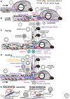
Figure 4.
Figure 4.
Autogenous origin of cilia with successive origins of different ciliary components under three contrasting selective forces. (A) During or just after the origin of nuclear pore complexes (npc Fig. 2), singlet microtubules from the γ-tubulin protocentrosome push out the plasma membrane as a protocilium by which plus-end-directed kinesin-2 motors (Verhey et al. 2011) attached by their tails to glycoprotein surface adhesins sticking to the substratum propel its microtubules forward (arrow) in primitive gliding motility, enabling cells to find fresh prey on the substratum as phagotrophy locally depletes it. (B) Posterior ciliary gliding was improved by attaching microtubules firmly to the cell surface by protocentriolar transitional fibers (proximally) and Y-shaped membrane connectors (slightly distally) plus ciliary compartmentation dependent on novel diffusion barriers (septin filaments in ciliary membrane base: Carvalho-Santos et al. 2011), central distal transition plate (TP), and a peripheral dense collar at the distal end of the Y-connector region, associated with npc proteins with novel ciliary import machinery using npcs and importin-β2 and NLS-related CLS targeting sequences (located in TP and/or collar). Dynein 1b, the first dynein, evolved (by gene-duplicating its common ancestor with the related AAA+ ATPase midasin/REA1 that mechanically strips biogenesis factors from 60S preribosomal subunits just before they exit the nucleus: Garbarino and Gibbons 2002; Kressler et al. 2012) to recycle distal adhesins left at the protociliary tip by axoneme gliding. Anterograde movement of dynein improved by carriage on kinesin-2-driven IFTB particles that evolved from CopI coat proteins, and special SNAREs evolved for basal delivery of ciliary membrane precursor vesicles with distinctive proteins and lipids. Protocentrioles were rigidified by a hub-spoke core, microtubules fixed at nine by ninefold SAS-6 hub assembly (Guichard et al. 2012). Cross-links rigidified the axoneme, the two on its substratum face having numerous extra-rigid linkers linking them like sled runners (subsequently becoming doublets 1 and 2 of Chlamydomonas: Lin et al. 2012). Novel proteins (e.g., Rib43a: Norrander et al. 2000) stabilized A-tubules and the bases of centriolar root microtubules (dorsal and ventral fans) compared with transient spindle microtubules. (C) Ciliary duplication produced a younger “fishing” cilium projecting into the medium for trapping swimming bacteria, pulling them baseward by minus-directed dynein 1b, for phagocytosis. Centrin plus novel proteins orthogonally rigidly connected the two protocentrioles now with doublets (architecture [Nicastro et al. 2011] perhaps determined by novel microtubule inner proteins, for example PACRG (Ikeda et al. 2007; Ikeda 2008), stabilized by a scaffold containing ε-tubulin [blue]). PACRG-interacting Rib72 (Ikeda et al. 2003) differentiated ciliary from centriolar doublets. Centriolar transformation temporally and physiologically differentiated the two cilia and made separate left and right ventral microtubular roots. (D) Successive dynein duplications generated inner arms (top: sufficient for bending [Heiss et al. 2013] as doublet 1–2 linker excluded arms from doublet 1, destroying ninefold symmetry; then the nexin–dynein regulatory complex [Heuser et al. 2009, 2012; Lin et al. 2011; Bower et al. 2013] for calcium regulation of beat; then outer dynein arms for greater power, and the center pair [nucleated by γ-tubulin on TP, and fixed so did not rotate] with new arms [Carbajal-Gonzalez et al. 2013] and kinesin-9 [Wickstead et al. 2010] and spokes [Barber et al. 2012] to modulate beat mechanics [not inherently needed for planar beat: Idei et al. 2013]) to draw in more prey by water currents. For pictorial simplicity, the cell body is proportionally too small and ahead of the cilia, but probably at stage b/c the cytoskeleton geometrically rearranged to lift the cell body from the substratum and put the ciliated cell apex at the front, which is mechanically stabler (found in all extant posterior ciliary gliding eukaryotes), entailing a basal stable bend ([E] mechanism unknown) for the posterior gliding cilium. Many complexities of present cilia (Mizuno et al. 2012) probably evolved subsequently to improve efficiency but would not have been essential for their origin, for example, association of IFTA/B into one complex and of these into distinct anterograde and retrograde trains (Pigino et al. 2009; Buisson et al. 2013), and addition of BBsomes, likely adaptors for improving retrograde transport of some proteins (Lechtreck et al. 2013) and sensory functions (sensation [Jékely and Arendt 2006] is less plausible than gliding for the original function), and beat pattern modulators. (E) The cenancestral eukaryote diverged to form swimming excavates that abandoned gliding and undulate the posterior cilium to draw prey into the ventral groove supported by a split right ventral centriolar root R2 (blue), and Euglenozoa that ancestrally retained gliding, added a cytopharnyx supported by ancestrally unsplit R2 and dissimilar paraxonemal rods (probably attached to the specially linked doublet 1–2 homologs) to broaden and further rigidify the posterior cilium for stabler gliding, and parallelized their centrioles within a ciliary pocket. After losing phagotrophy, saprophytic, parasitic, and photosynthetic Euglenozoa lost gliding and developed swimming by the anterior (Euglenophyceae) or posterior (trypanosomatids) cilium; some bacterivores (petalomonads) lost the posterior cilium, presumably recruiting dynein 1b for anterior ciliary gliding. All other eukaryotes evolved from excavates (Fig. 3); Apusozoa, Cercozoa, and the heterokont Caecitellus reevolved posterior ciliary gliding, presumably using kinesin-2. The V-fiber, with associated acorn-base attached distally to centriole 1–2 triplets (at least in neokaryotes: Geimer and Melkonian 2005), demarcates the centriole from the transition zone and perhaps evolved in the cenancestral eukaryote; its rotational asymmetry and that of centriolar root attachments to specific triplets probably reflect an asymmetric doublet “numbering machinery” that probably evolved in the earliest gliding protocilium (B).
Figure 5.
Figure 5.
Eukaryote cell-cycle logic and evolution. Complexes of cyclin-dependent kinase (CDK) and cyclins control the eukaryotic cell cycle by phosphorylating numerous proteins, timed by growth-dependent increases in cyclins and their sudden proteolysis (red curves) (Nasmyth 1995). Cyclins share a domain with neomuran-specific transcription factor TFII, and CDKs evolved from posibacterial serine-threonine (S/T) kinases. Originally one cyclin could have controlled S-phase initiation and anaphase onset using a lower threshold for the former switch (Novak et al. 1998; Tyson and Novak 2008; Harashima et al. 2013); gene duplication enabled better control by cyclinE/cdk2 (for DNA replication and centriole duplication) and cyclin B/cdk1 to activate the anaphase promoting complex (APC), the ubiquitin ligase that initiates anaphase resetting of cell-cycle controls via proteasome degradation of cyclins and numerous key cycle proteins. Phosphorylation-cum-proteolytic cell-cycle controls originated in posibacteria, but a novel Cdc-6-mediated control over replication initiation evolved in ancestral neomura after histones H3/4 and MCM DNA helicase, replacing eubacterial DnaA/CrtA control; proteolysis by proteasomes that originated in the neomuran/actinobacterial cenancestor replaced eubacterial ClpXP proteolysis. Ancestral eukaryotes evolved origin recognition complexes (ORCs) more complex than the single protein Cdc-6 of archaebacteria, probably because the suddenness of mitotic anaphase (rapidly segregating all parts of chromosomes at once, unlike the temporally separate segregation of origins and the generally single terminus in bacteria) required concerted replication initiation at hundreds of origins, ensuring replication completion and tighter chromosome folding (using extra histones and novel heterochromatin machinery: Cavalier-Smith 2010c) well before mitosis; uniquely in eukaryotes, mitosis demands a temporally discrete S phase. Formerly, only neokaryotes were thought to have ORCs (Cavalier-Smith 2010b), but extremely divergent versions of most constituents are now known in trypanosomes, whose cell-cycle controls are the most divergent within eukaryotes (Li 2012), consistent with eukaryotic rooting between Euglenozoa and neokaryotes (Figs. 1 and 3). Successively more complex controls and checkpoints evolved with novel polo-like and aurora S/T kinases playing multifarious roles in mitosis and cytokinesis and a multiplicity of kinesins evolving to improve spindle assembly and function. Probably all proteins shared by trypanosomes (Li 2012) and opisthokonts evolved before the eukaryote cenancestor. Mitosis (upper panel) was converted to meiosis by two innovations (lower panel): homologous chromosome pairing by the synaptonemal complex and blocking centromeric cohesin digestion at meiosis 1 anaphase, which automatically bypassed cell-cycle resetting caused by anaphase centromere splitting so that the next interphase had no S phase as previously explained (Cavalier-Smith 1981), thereby halving ploidy—the original meiotic function (Cavalier-Smith 2002a, 2010c). It is unclear whether centromeres evolved from posibacterial plasmid partition proteins associated with GTPase TubZ (Aylett et al. 2010, 2013) or de novo; CENP-A was probably added only in neokaryotes after they diverged from Euglenozoa (Fig. 3) (Cavalier-Smith 2010c). As the text notes, recent evidence suggests that cohesin digestion is not invariably necessary for centriole separation in animals, raising doubt as to the ancestral eukaryotic process.

References

    1. Adams DW, Errington J 2009. Bacterial cell division: Assembly, maintenance and disassembly of the Z ring. Nat Rev Microbiol 7: 642–653 - PubMed
    1. Aylett CH, Wang Q, Michie KA, Amos LA, Lowe J 2010. Filament structure of bacterial tubulin homologue TubZ. Proc Natl Acad Sci 107: 19766–19771 - PMC - PubMed
    1. Aylett CH, Izore T, Amos LA, Lowe J 2013. Structure of the tubulin/FtsZ-like protein TubZ from Pseudomonas bacteriophage ϕKZ. J Mol Biol 425: 2164–2173 - PMC - PubMed
    1. Bapteste E, Susko E, Leigh J, Ruiz-Trillo I, Bucknam J, Doolittle WF 2008. Alternative methods for concatenation of core genes indicate a lack of resolution in deep nodes of the prokaryotic phylogeny. Mol Biol Evol 25: 83–91 - PubMed
    1. Barber CF, Heuser T, Carbajal-Gonzalez BI, Botchkarev VV Jr, Nicastro D 2012. Three-dimensional structure of the radial spokes reveals heterogeneity and interactions with dyneins in Chlamydomonas flagella. Mol Biol Cell 23: 111–120 - PMC - PubMed

LinkOut - more resources