

Bilateral congenital ureteral strictures in a young cat
- PMID: 25183890
- PMCID: PMC4137923
Bilateral congenital ureteral strictures in a young cat
Abstract
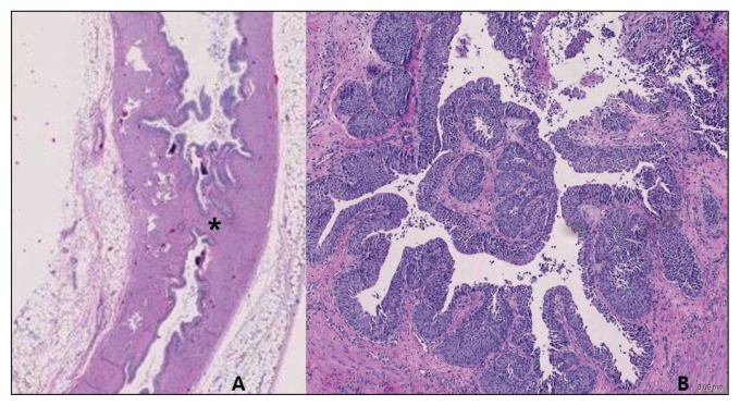
An 8-month-old cat was presented with bilateral hydronephrosis. Bilateral ureteral obstructions were identified by diagnostic imaging and confirmed by necropsy. Histopathologic findings revealed polypoid transitional epithelial hyperplasia with chronic lymphoplasmacytic inflammation. This report documents congenital ureteral strictures as a cause of ureteral obstruction in a young cat.
Constriction urétérale bilatérale congénitale chez un jeune chat. Un chat âgé de 8 mois a été présenté avec une hydrophénose bilatérale. Des obstructions urétérales bilatérales ont été identifiées par imagerie diagnostique et confirmée par nécropsie. Les résultats histopathologiques ont révélé une hyperplasie épithéliale polypoïde transitionnelle avec une inflammation lymphoplasmacytique chronique. Ce rapport documente les constrictions urétérales congénitales comme cause de l’obstruction urétérale chez un jeune chat.(Traduit par Isabelle Vallières).
Figures


References
-
- Hardie EM, Kyles AE. Management of ureteral obstruction. Vet Clin Small Anim. 2004;34:989–1010. - PubMed
-
- Kyles AE, Hardie EM, Wooden BG, et al. Clinical, clinicopathologic radiographic, and ultrasonographic abnormalities in cats with ureteral calculi: 163 cases (1984–2002) J Am Vet Med Assoc. 2005;226:932–936. - PubMed
-
- Kyles AE, Hardie EM, Wooden BG, et al. Management and outcome of cats with ureteral calculi: 153 cases (1984–2002) J Am Vet Med Assoc. 2005;226:937–944. - PubMed
-
- Westropp JL, Ruby AL, Bailiff NL, et al. Dried solidified blood calculi in the urinary tract of cats. J Vet Intern Med. 20:828–834. - PubMed
-
- Zaid MS, Berent AC, Weisse C, Caceres A. Feline ureteral strictures: 10 cases (2007–2009) J Vet Intern Med. 2011;25:222–229. - PubMed
Publication types
MeSH terms
LinkOut - more resources
Full Text Sources
Miscellaneous