

Quantitative analysis of bone and soft tissue by micro-computed tomography: applications to ex vivo and in vivo studies
- PMID: 25184037
- PMCID: PMC4140449
- DOI: 10.1038/bonekey.2014.59
Quantitative analysis of bone and soft tissue by micro-computed tomography: applications to ex vivo and in vivo studies
Abstract
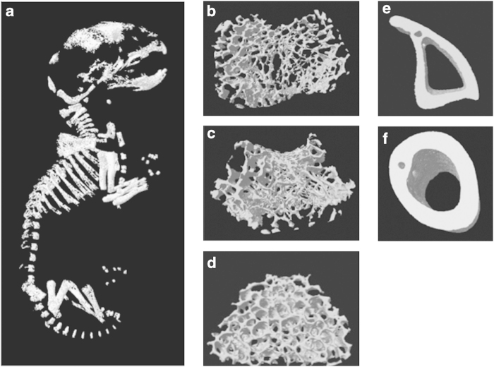
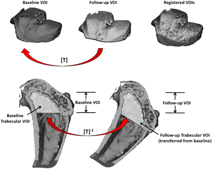
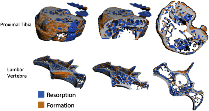
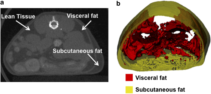
Micro-computed tomography (micro-CT) is a high-resolution imaging modality that is capable of analysing bone structure with a voxel size on the order of 10 μm. With the development of in vivo micro-CT, where disease progression and treatment can be monitored in a living animal over a period of time, this modality has become a standard tool for preclinical assessment of bone architecture during disease progression and treatment. For meaningful comparison between micro-CT studies, it is essential that the same parameters for data acquisition and analysis methods be used. This protocol outlines the common procedures that are currently used for sample preparation, scanning, reconstruction and analysis in micro-CT studies. Scan and analysis methods for trabecular and cortical bone are covered for the femur, tibia, vertebra and the full neonate body of small rodents. The analysis procedures using the software provided by ScancoMedical and Bruker are discussed, and the routinely used bone architectural parameters are outlined. This protocol also provides a section dedicated to in vivo scanning and analysis, which covers the topics of anaesthesia, radiation dose and image registration. Because of the expanding research using micro-CT to study other skeletal sites, as well as soft tissues, we also provide a review of current techniques to examine the skull and mandible, adipose tissue, vasculature, tumour severity and cartilage. Lists of recommended further reading and literature references are included to provide the reader with more detail on the methods described.
Figures
Similar articles
-
The effect of voxel size on high-resolution peripheral computed tomography measurements of trabecular and cortical bone microstructure.Med Phys. 2012 Apr;39(4):1893-903. doi: 10.1118/1.3689813. Med Phys. 2012. PMID: 22482611 Free PMC article.
-
Comparison of ex vivo and in vivo micro-computed tomography of rat tibia at different scanning settings.J Orthop Res. 2017 Aug;35(8):1690-1698. doi: 10.1002/jor.23435. Epub 2016 Oct 12. J Orthop Res. 2017. PMID: 27626898
-
A comparative evaluation of cone beam CT and micro-CT on trabecular bone structures in the human mandible.Dentomaxillofac Radiol. 2013;42(8):20130145. doi: 10.1259/dmfr.20130145. Epub 2013 Jul 5. Dentomaxillofac Radiol. 2013. PMID: 23833320 Free PMC article.
-
High-resolution computed tomography for clinical imaging of bone microarchitecture.Clin Orthop Relat Res. 2011 Aug;469(8):2179-93. doi: 10.1007/s11999-010-1766-x. Clin Orthop Relat Res. 2011. PMID: 21344275 Free PMC article. Review.
-
Micro-CT analysis of the rodent jaw bone micro-architecture: A systematic review.Bone Rep. 2015 Jan 21;2:14-24. doi: 10.1016/j.bonr.2014.10.005. eCollection 2015 Jun. Bone Rep. 2015. PMID: 28525530 Free PMC article. Review.
Cited by
-
A method to quantify and visualize femoral head intraosseous arteries by micro-CT.J Anat. 2016 Aug;229(2):326-33. doi: 10.1111/joa.12475. Epub 2016 Apr 14. J Anat. 2016. PMID: 27074892 Free PMC article.
-
Quantitative in vivo micro-computed tomography for monitoring disease activity and treatment response in a collagen-induced arthritis mouse model.Sci Rep. 2022 Feb 21;12(1):2863. doi: 10.1038/s41598-022-06837-w. Sci Rep. 2022. PMID: 35190580 Free PMC article.
-
Three-Dimensional Ultrasound Versus Computerized Tomography in Fat Graft Volumetric Analysis.Ann Plast Surg. 2018 Mar;80(3):293-296. doi: 10.1097/SAP.0000000000001183. Ann Plast Surg. 2018. PMID: 28678028 Free PMC article.
-
Efficacy of photobiomodulation on accelerating bone healing after tooth extraction: a systematic review.Lasers Med Sci. 2019 Jun;34(4):685-692. doi: 10.1007/s10103-018-2641-3. Epub 2018 Oct 11. Lasers Med Sci. 2019. PMID: 30311084
-
Bidirectional regulation of bone formation by exogenous and osteosarcoma-derived Sema3A.Sci Rep. 2018 May 2;8(1):6877. doi: 10.1038/s41598-018-25290-2. Sci Rep. 2018. PMID: 29720701 Free PMC article.
References
-
- Turner CH, Burr DB. Basic biomechanical measurements of bone: a tutorial. Bone 1993;14:595–608. - PubMed
-
- van 't Hof RJ. Analysis of bone architecture in rodents using microcomputed tomography. Methods Mol Biol 2012;816:461–476. - PubMed
-
- Bouxsein ML, Boyd SK, Christiansen BA, Guldberg RE, Jepsen KJ, Müller R. Guidelines for assessment of bone microstructure in rodents using micro-computed tomography. J Bone Miner Res 2010;25:1468–1486. - PubMed
-
- Stauber M, Müller R. Micro-computed tomography: a method for the non-destructive evaluation of the three-dimensional structure of biological specimens. Methods Mol Biol 2008;455:273–292. - PubMed
LinkOut - more resources
Full Text Sources
Other Literature Sources