. 2014 Jul;3(3):179-90.
doi: 10.4103/2303-9027.138793.

Techniques of imaging of nodal stations of gastric cancer by endoscopic ultrasound

Affiliations

Techniques of imaging of nodal stations of gastric cancer by endoscopic ultrasound

Malay Sharma et al. Endosc Ultrasound. 2014 Jul.

Abstract

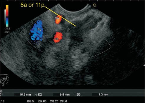
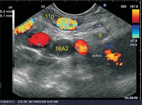
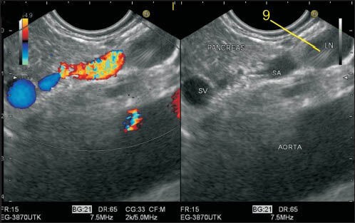
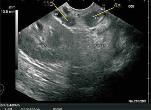
Nodal staging is of crucial importance in the management of gastric cancer (GC). The available modalities of nodal imaging in GC do not provide a high sensitivity and specificity of lymph node status. Comparative study of endoscopic ultrasonography (EUS) and multislice spiral computed tomography in GC has shown greater accuracy of EUS for N staging. EUS is not used routinely in patients with GC as it is not available at all centers, and its accuracy is operator dependent. Standard techniques of identification of nodal station (as suggested by Japanese Research Society for the Study of Gastric Cancer) by EUS have not been described so far. Identification of each nodal station by EUS requires adequate knowledge of anatomy as well as understanding the proper technique to perform EUS. This review presents a method to identify the regional nodal stations of GC by linear EUS and hence will help in appropriate N staging of GC.

Keywords: Endoscopic ultrasound; gastric cancer; lymph node; multidetector spiral computed tomography.


Conflict of interest statement

Conflict of Interest: None declared.

Figures

Figure 1a
Figure 1a
This figure shows lymph nodes near right paracardial area, station 1; lesser curvature, station 3; supra pyloric area, station 5; left paracardial area, station 2; greater curvature, station 4; and infrapyloric area station 6
Figure 1b
Figure 1b
This figure shows N2 stations near; left gastric artery (station 7), common hepatic artery (station 8), celiac artery (station 9) and splenic artery (stations 10 and 11). Lymph nodes (LNs) of N3 stations are seen near hepatoduodenal ligament (station 12), the posterior aspect of the pancreas (station 13), the root of the mesentery (station 14), the transverse mesocolon (station 15) and para-aortic area (station 16). The para-aortic LN at station 16 are seen above (16a) and below (16b) the lower border of left renal vein where it crosses the aorta. Additional stations (17-20) include lower thoracic, paraesophageal and diaphragmatic nodes
Figure 2
Figure 2
The gastroesophageal (GE) junction lies about 3-4 cm below the diaphragm. The GE junction demarcates the area between the paracardial stations (stations 1 and 2) and the lesser and greater curvature stations (stations 3 and 4). The lymph node (LN) of station 1 lies in the right paracardial area, and the LN of station 2 lie in left paracardial area. The LN in this figure belongs to station 1 and lies between the wall of the stomach and the left lobe of the liver and lies along esophageal branch of left gastric artery. An LN belonging to station 7 is also seen. LGA: Left gastric artery, SA: Splenic artery
Figure 3
Figure 3
The Lymph node (LN) of station 1 lies on the right side of esophagogastric junction. This LN lies between the wall of the stomach and the left lobe of the liver in the hepatogastric ligament, which lies between the liver and stomach. Two LNs belonging to station 7 and 11p are also seen. The identification of hepatogastric ligament is done as the hyperechoeic area between liver and stomach during anticlockwise rotation after visualizing the aorta
Figure 4
Figure 4
The imaging of the left paracardial and right paracardial areas can be done along a plane which lies posteriorly along the anterior border of aorta and anteriorly goes through the segments 2 and 3 of the left lobe of the liver. Initially, the segments 2 and 3 of liver are visualized in an open position by echoendoscope. A clockwise rotation of the echoendoscope from this position traces the right paracardial area till it reaches the hilum of the liver and traces it on further rotation till the aorta is visualized after a 180° rotation. An anticlockwise rotation from segments 2 and 3 of liver traces the left paracardial area where the origin of left inferior phrenic artery is seen as the first branch from the anterolateral surface of the abdominal aorta above the origin of the celiac artery. The gastrophrenic ligament lies in left paracardial area where lymph node of station 2 lies in at the left margin of the gastroesophageal junction along the esophagocardiac branches of left inferior phrenic artery
Figure 5a
Figure 5a
The lesser curvature of the stomach is approximately 10 cm in distance and can be divided into two parts. The branches of left gastric artery lie in the upper part of the lesser curvature in the hepatogastric ligament near the upper part of the lesser curvature (about 5 cm below the esophagogastric junction). The station 3 lymph nodes (LNs) lie along the lesser curvature of the stomach. The LNs up to 5 cm below the junction are included in the upper part of the lesser curvature. CAL: Celiac artery
Figure 5b
Figure 5b
In this image, the left gastric vein is seen joining the upper part of the portal vein (PV) and this lymph node (LN) lies very close to the upper part of the lesser curvature. The left gastric artery and vein run together in the hepatogastric ligament and a LN, which lies close to either, can be included in 3a. The LN labeled as 11p lies more close to the splenic artery than to the lesser curvature and hence is not included in station 3a. An LN lying near the PV (12p) is also seen. The LNs of the 3b (not seen in this image) station lie along the right gastric artery in the lower part of the lesser curvature. The origin and course of right gastric artery is difficult to identify on endoscopic ultrasonography. LGV: Left gastric vein, PV: Portal vein
Figure 6
Figure 6
The lienorenal ligament is seen as a structure between the splenic hilum and the left kidney and includes the tail of pancreas. In this ligament the splenic artery and splenic vein traverse toward the hilum of the spleen but the lymph node (LN) is away from the vessels. In this case, a LN lying at the hilum of the spleen close to the tail of PAN is seen and belongs to station 4. LK: Left kidney, D1, D2: Diameter of node
Figure 7
Figure 7
The station 3 lymph nodes (LNs) lie along the lower part of the lesser curvature of the stomach where the pancreas is also seen. This LN belongs to station 3b as it lies close to the lower part of the lesser curvature. In this image the portal vein (PV) is seen behind the PAN and an LN of station 12p lies close to PV. An LN lying behind inferior vena cava belonging to retrocaval group is also seen. RRA: Right renal artery, HOP: Head of pancreas
Figure 8
Figure 8
In this image, the LN lies at the upper border of the pancreas between the lesser curvature and the left gastric vein (LGV). The LGV is seen joining the upper border of portal vein (PV) near the portal venous confluence. The splenic artery (SA) is seen. This lymph node (LN) is in proximity of two vessels; the PV and SA. However, this LN lies very close to the lower part of the lesser curvature of the stomach and belongs to station 3b. PVC: Portal venous confluence
Figure 9
Figure 9
Two lymph nodes are seen adjacent to the greater curvature. They belong to station 4. It is difficult to identify the blood vessels along the greater curvature because of their smaller caliber
Figure 10
Figure 10
Imaging from the duodenal bulb can show the portal vein (PV) and the bifurcation of common hepatic artery into the hepatic artery proper and gastroduodenal artery. In this image, the lymph node is seen between the supra pancreatic part of PV and the wall of the duodenum and belongs to station 5
Figure 11
Figure 11
Imaging from the duodenal bulb can also show the portal vein and the bifurcation of common hepatic artery into the hepatic artery (HA) proper and gastroduodenal artery. The HA tends to run in a cranial direction toward the hilum of the liver and the gastroduodenal artery tends to go down behind the duodenum. In this image twolymph nodes (LNs) are seen and station 5 LN lies between duodenum and liver while station 6 LN lies between duodenum and PAN. A LN of station 12p is also seen. PAN: Pancreas, CHA: Common hepatic artery
Figure 12
Figure 12
The lymph node (LN) labeled as station 7 lies a little away from the wall of stomach and close to the trunk of left gastric artery (LGA). The origin of LGA from the celiac artery is seen. The other LN belongs to station 11p. Sometimes it is possible to follow the branches of LGA to the level of the origin of esophageal branches
Figure 13
Figure 13
The lymph node (LN) labeled as station 8a lies close to the common hepatic artery. One LN lies anterior to hepatic artery (HA), and one LN (8p) lies anterior to inferior vena cava posterior to HA. The LN labeled as 8a can be also included in station 3b and such cases the proximity to the vessel or to the wall of the stomach will decide the station. In this case, the station 8a LN is equidistant from HA and from wall of the stomach. HA: Hepatic artery
Figure 14
Figure 14
The lymph node (LN) labeled as station 8p lies close to the common hepatic artery. This LN lies anterior to inferior vena cava and posterior to hepatic artery
Figure 15
Figure 15
This lymph node (LN) cluster is seen anterior to hepatic artery (HA) as well as near the proximal part of the splenic artery (SA). One of the LNs is touching the SA and can be labeled as either 8a or 11p. At this point, the distance between HA and SA is only about 5 mm, and it is not possible to differentiate between 8a and 11p
Figure 16
Figure 16
The lymph node (LN) labeled as station 9 lies just above the celiac artery (CAL) and in front of the abdominal aorta. The CAL takes a course anteriorly after taking origin from the abdominal aorta. This LN is seen just above the anterior most part of CAL. The trunk of left gastric artery near its origin is also seen, but this LN is closest to the CAL and hence is labeled as station 9. Another LN lying below the CAL lies close to the abdominal aorta and belongs to station 16A2. A LN belonging to 11p station lies near the lower border of the splenic artery
Figure 17
Figure 17
The celiac artery (CAL) takes a vertical course anteriorly after taking origin from the abdominal aorta. This lymph node (LN) is seen just above the anterior most part of CAL, which is not seen in this frame. The splenic artery (SA) is the most tortuous artery of the whole body and in this frame is seen lying anterior to aorta. The LN lying above SA is closer to CAL and anterior to aorta and thus belongs to station 9
Figure 18
Figure 18
The splenic artery (SA) runs a tortuous course and sometimes dips behind and above the pancreas. In this image, two lymph node (LN) are seen, and the LN labeled as 11d is seen close to the distal part of SA near the upper border of the body of PAN. The upper LN is at station 4
Figure 19
Figure 19
This image shows another lymph node (LN) in the lienorenal ligament close to the hilum of the spleen and can be included either in station 11d or 10. The station 10 LN lies more close to the pancreatic tail whereas station 11d lies near the upper border of the body of pancreas. On endoscopic ultrasonography a demarcation of the point where the body of PAN becomes tail can be seen when the renal artery and renal vein are no longer visualized because of the entry into to the hilum of the left kidney. So the inclusion of this LN can be done in station 10
Figure 20
Figure 20
In this case one-lymph node (LN) labeled as 11p lies above the splenic artery as it takes a tortuous course and dips down. The LN labeled as 12p lies in the hepatoduodenal ligament just above the upper border of head of the pancreas as the portal vein exits near the upper border on its way to the hilum of the liver
Figure 21
Figure 21
The portal vein (PV) near its formation lies anterior to the inferior vena cava (IVC) and maintains this position till it reaches the hilum. In this case the lymph node (LN) labeled as 12p lies posterior to PV and anterior to IVC. The location of this LN can be also included as precaval because it lies on the anterior surface of the proximal half of IVC. This LN is equidistant from both vessels. The retrocaval LN lies behind IVC anterior to the crux of the diaphragm. The LN of station 8a is also seen lying anterior to hepatic artery
Figure 22
Figure 22
The bifurcation of the celiac artery into the hepatic artery (HA) and splenic artery (SA) produces a vertical seagull like appearance with vascular signals on endoscopic ultrasonography from stomach where the limb coming toward the probe is SA and the limb going away from the probe is HA. In between the two vessels lies the highest point of pancreas, which is called tuber omentale
Figure 23
Figure 23
In this case a lymph node (LN) is seen just above the tuber omentale and is close to four vessels, which are celiac artery, hepatic artery, splenic artery and portal vein. This LN lies in the lower part of the gastro hepatic omentum and can be included in station 9, 11p, 8a and 12p
Figure 24
Figure 24
The lymph node labeled as 12b lies along the common bile duct in the curve below union of the cystic duct with gallbladder. This lies in the hepatoduodenal part of lesser omentum and is best visualized from the bulb. CBD: Common bile duct, GB: Gall bladder
Figure 25a
Figure 25a
The lymph node (LN) labeled as 13 lies behind the pancreas just close to the lower end papilla and anterior to inferior vena cava. The can be included in station 12b (close to common bile duct) or precaval LN also
Figure 25b
Figure 25b
In this case two-lymph nodes (LNs) are seen. Station 13 LN lies behind pancreas but lies away from the aorta. Station 16A2 lies below the celiac artery above the renal vein anterior to aorta
Figure 26
Figure 26
Imaging from stomach can sometimes push the scope below the level of head and body of pancreas. A lymph node (LN) lying cranial to duodenal papilla during imaging from 2nd part of the duodenum can be considered as station 13 whereas an LN lying below the papilla will lie close to the lower border of PAN and will belong to station 18
Figure 27
Figure 27
The superior mesenteric artery (SMA) is the second ventral branch of the aorta, given off slightly below the celiac trunk, which descends in a groove on the posterior surface of the neck of the pancreas. Below the inferior margin of the neck of the PAN, it goes anterior to the uncinate process and the horizontal portion of the duodenum and enters the root of the mesentery. It is easy to identify lymph node (LN) close to SMA and superior mesenteric vein (SMV) from the horizontal portion of the duodenum. In this image, the LN is seen near the root of mesentery close to the origin of SMA and also close to the place where the SMV is about to join splenic vein for formation of the portal vein. The SMA nodes (14a) can lie behind the body of PAN, on the uncinate process of PAN or near the root of mesentery
Figure 28
Figure 28
The station 14v nodes lie close to superior mesenteric vein
Figure 29a
Figure 29a
Within the mesentery, the main arterial stem of superior mesenteric artery (SMA) describes an arc that spans the distance between the horizontal duodenum and the ileocecal junction, where the SMA terminates by anastomosing with one of its own branches, the ileocolic artery. Of the arteries to the large intestine, the middle colic artery (MCA) is the first to arise from the right side of the SMA. This branch is usually given off at the inferior margin of the neck of the pancreas before the SMA enters the mesentery. The MCA passes into the transverse mesocolon. In this image, the jejunal branches are seen going away from the probe, and the inferior pancreaticoduodenal artery is seen as the first branch arising from the right side of the SMA coming toward the probe. The second branch coming from the right side of SMA is MCA, which is seen to enter the transverse mesocolon. MCA: Middle colic artery, INFR PDA: Inferior pancreaticoduodenal artery, IMV: Inferior mesenteric vein
Figure 29b
Figure 29b
The lymph node (LN) lying along middle colic artery belongs to station 15. In the second image, a LN is also seen along the first-jejunal vein
Figures 30a and b
Figures 30a and b
The middle colic artery (MCA) is seen taking origin from the right side of superior esenteric artery from the horizontal part of the duodenum. The course of the MCA is followed-up to the lymph node present in transverse mesocolon (station 15)
Figure 31
Figure 31
The distance between the esophageal hiatus and the aortic hiatus is about 1.5 cm, and the crux of diaphragm comes in between the two. A lymph node (LN) lying between the esophageal and aortic hiatus can be differentiated by the location: A LN between crux and aorta will be called as aortic hiatus node (16A1) whereas an LN between esophageal hiatus and crux will be called as esophageal hiatal node belonging to station 20
Figure 32
Figure 32
This lymph node (LN) belonging to station 16A2 lies below the celiac artery and posterior to the origin of superior mesenteric artery (SMA). Although this LN is very close to the posterior surface of SMA, the LN lies anterior to aorta and this LN is not included in the root of mesentery LN (station 14a)
Figure 33
Figure 33
In this image, the upper and lower boundary of station A2 are seen as the upper border of the celiac artery (CAL) (orange line) and lower border of left renal vein (LRV) (blue line). The lymph node (LN) belonging to station 16A2 lies below the CAL and above the lower margin of LRV anterior to the aorta. Although the LNs lie posterior to the pancreas, the location near the aorta places them in para-aortic group
Figure 34
Figure 34
The lower border of left renal vein (LRV) demarcates the upper border of station 16B1. This image shows a lymph node anterior to aorta and below the lower border of LRV belonging to station 16B1. The lower border of station 16B1 is identified by origin of inferior mesenteric artery, which is not seen in these images. LRV: Left renal vein
Figure 35
Figure 35
This image shows 16B1 lymph node in a different patient
Figure 36
Figure 36
In this case two-lymph nodes (LNs) are seen: One LN lies above the origin of inferior mesenteric artery from the aorta, and the second LN lies below the level of the upper margin of origin of IMA. The upper LN belongs to station 16B1, and the lower LN belongs to station 16B2. It is uncommon to see the origin of IMA from aorta by endoscopic ultrasonography but in this image the LNs can be seen from the stomach below the level of the pancreas. IMA: Inferior mesenteric artery
Figure 37
Figure 37
The fine-needle aspiration cytology was done from the 16B1station
Figure 38
Figure 38
Imaging from stomach can sometimes push the scope below the level of head and body of pancreas. A lymph node (LN) lying cranial to duodenal papilla can be considered as station 13 whereas a LN lying below the papilla will lie near the lower border of PAN and will belong to station 18. In this case, the papilla is not seen but two LN lies behind the PAN in retroperitoneum and belongs to station 13 and another LN, which belong to station 18 lies along the inferior border of the body of PAN
Figure 39
Figure 39
The station 18 lymph nodes (LNs) are retroperitoneal and lie along the inferior border of the body of pancreas. In this case, the LN is seen from the horizontal part of duodenum anterior to the superior mesenteric vein (SMV) just below the place where it crosses the uncinate process. The part of PAN lying in front of SMV belongs to the uncinate process
Figure 40
Figure 40
A color Doppler shows the superior mesenteric vein and the lymph node belonging to station 18 from the horizontal part of the duodenum
Figure 41
Figure 41
In this case the station 19 and 20 lymph node (LN) are seen. The station 19 LN is retroperitoneal and lies on the inferior surface of the diaphragm. In this case, the LN is much away from the esophageal hiatus and also away from the aorta. The diaphragm is not seen in this image, but the location anterior to crux, below the esophageal hiatus suggest this LN as station 19. The station 20 LN lies between the esophageal hiatus and crux of diaphragm
Figure 42
Figure 42
The lymph node (LN) in supra diaphragmatic location lie near the esophagus: Paraesophageal below the pulmonary vein (110) (a), below the left atrium (LA) (111) (b), close to inferior vena cava (supra diaphragmatic) (c) and close to aorta in a posterior mediastinal location (112) (d). The International Association for the Study of Lung Cancer classification of lung cancer staging identifies these LNs in a different manner as station 8 and station 9 LN. RIPV: Right inferior pulmonary vein, ST7: Station 7 of international association of study of lung cancer classification for LN. LA: Left atrium, RA: Right atrium

References

    1. Crew KD, Neugut AI. Epidemiology of gastric cancer. World J Gastroenterol. 2006;12:354–62. - PMC - PubMed
    1. Park SR, Kim MJ, Ryu KW, et al. Prognostic value of preoperative clinical staging assessed by computed tomography in resectable gastric cancer patients: A viewpoint in the era of preoperative treatment. Ann Surg. 2010;251:428–35. - PubMed
    1. Jeong JY, Kim MG, Ha TK, et al. Prognostic factors on overall survival in lymph node negative gastric cancer patients who underwent curative resection. J Gastric Cancer. 2012;12:210–6. - PMC - PubMed
    1. Japanese Gastric Cancer Association. Japanese classification of gastric carcinoma: 3rd English edition. Gastric Cancer. 2011;14:101–12. - PubMed
    1. Kajitani T. The general rules for the gastric cancer study in surgery and pathology. Part I. Clinical classification. Jpn J Surg. 1981;11:127–39. - PubMed