Review
. 2014 Sep;78(3):418-37.
doi: 10.1128/MMBR.00012-14.

Picornavirus morphogenesis

Affiliations
Review

Picornavirus morphogenesis

Ping Jiang et al. Microbiol Mol Biol Rev. 2014 Sep.

Abstract

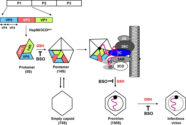
The Picornaviridae represent a large family of small plus-strand RNA viruses that cause a bewildering array of important human and animal diseases. Morphogenesis is the least-understood step in the life cycle of these viruses, and this process is difficult to study because encapsidation is tightly coupled to genome translation and RNA replication. Although the basic steps of assembly have been known for some time, very few details are available about the mechanism and factors that regulate this process. Most of the information available has been derived from studies of enteroviruses, in particular poliovirus, where recent evidence has shown that, surprisingly, the specificity of encapsidation is governed by a viral protein-protein interaction that does not involve an RNA packaging signal. In this review, we make an attempt to summarize what is currently known about the following topics: (i) encapsidation intermediates, (ii) the specificity of encapsidation (iii), viral and cellular factors that are required for encapsidation, (iv) inhibitors of encapsidation, and (v) a model of enterovirus encapsidation. Finally, we compare some features of picornavirus morphogenesis with those of other plus-strand RNA viruses.


Figures

FIG 1
FIG 1
Structure of poliovirus. (A) Schematic diagram of the structure of poliovirus with icosahedral symmetry (3, 219). The 5-fold, 3-fold, and 2-fold axes of symmetry are indicated. The capsid proteins VP1 (blue), VP2 (yellow), and VP3 (magenta) make up the outer surface of the particle, whereas VP4 is located internally. The structure shown in color is the processed protomer of which VP0 has already been cleaved into VP4 and VP2. The canyon around the 5-fold axis of symmetry is indicated with a ring. (Modified from reference with permission of the publisher. Copyright 1989 Annual Reviews.) (B) Computer model of poliovirus. The 5-fold, 3-fold, and 2-fold axes of symmetry and the canyon are visible on the structure. (Reprinted from reference with permission.) (C) Schematic representation of the three large poliovirus capsid proteins, each of which forms an eight-stranded, wedge-like, antiparallel β-barrel core (a) (3, 219). The antiparallel strands are connected by loops (BC, HI, DE, FG, GH, and CD). In panels b to d, the large capsid proteins are represented with ribbon diagrams (219). The four major neutralization antigenic sites (N-Ags) of poliovirus (type 1) map to surface loop extensions, as shown. N-AgI is a linear antigenic site that maps to the BC loop (amino acids 95 to 105) of VP1. All other major sites are discontinuous in nature: N-AgII (dotted line) spans VP1 and VP2 (amino acids 221 to 226 of VP1 and amino acids 164 to 172 of VP2). N-AgIII presents as two independent sites: N-AgIIIA consists of amino acids 58 to 60 and 71 to 73 of VP3 (dotted line), whereas N-AgIIIB contains amino acids 72 of VP2 and 76 to 79 of VP3. (Adapted from reference with permission of AAAS.) (D) Localization of all major neutralization antigenic sites on the poliovirion indicating the density of possible neutralizing antibody-binding sites. (a) Band diagram of a pentamer containing the apex of the 5-fold symmetry axis. N-Ags are shown as white balls surrounding the mesa. Binding of antibodies to N-AgI, located on the rim of the canyon, leads to the neutralization of the virus by preventing attachment of the virus to the cellular receptor. (b) Dense distribution of N-Ags throughout the poliovirus capsid. (c to e) Band diagrams and neutralization antigenic sites of the capsid proteins VP1, VP2, and VP3. (Reprinted from reference with permission of the publisher.)
FIG 2
FIG 2
Genome structure of picornavirus and polyprotein processing. The picornavirus genome is a linear single-strand RNA ranging from 7.1 to 8.9 kb in length. The RNA has a covalently linked viral protein (VPg) at its 5′ end and poly(A) at its 3′ end. The ORF is flanked by a long 5′ nontranslated region (NTR) and a short 3′ NTR. The long 5′ NTR contains an internal ribosome entry site (IRES) that directs polyprotein translation. Besides the highly structured 5′ NTR and 3′ NTR, other essential RNA secondary structures have been identified in the open reading frame. For poliovirus, these structures are the cis replication element (cre) located in the 2CATPase coding sequence (106) and two RNA elements, α and β, located in the 3Dpol coding region (110). All known picornavirus genomes contain a cre element, but these structures may map to different locations in the genomes of different viruses. The first cre discovered maps to the P1 coding region of human rhinovirus 14 (HRV14) (174). No analyses of α and β elements in picornavirus genomes other than poliovirus have been published. The single open reading frame is organized as 1ABCD-2ABC-3ABCD, with the numbers indicating the three different domains and each letter representing a protein. The P1 region encodes the capsid structural polypeptides. The P2 and P3 regions encode the nonstructural proteins associated with replication. The polyproteins of member viruses of a large number of picornavirus genera (Aphthovirus, Erbovirus, Kobuvirus, Cardiovirus, Teschovirus, Sapelovirus, and Senecavirus) other than Enterovirus or Hepatovirus have an additional protein, the L protein, attached to the N terminus (8).
FIG 3
FIG 3
Genome organization of poliovirus (PV) and hepatitis A virus (HAV) and steps in virion formation. The general PV/HAV genome organization is the same: a VPg-linked genome; a long 5′ NTR harboring replication signals and the IRES; a single ORF divided into P1 (capsid region [green]) and P2 plus P3, encoding nonstructural proteins; and 3′-terminal RNA structures terminated by poly(A). (A) PV encodes two proteinases (red and orange), of which 3Cpro functions mostly as a processing precursor of 3CDpro. The proteinase 2Apro cleaves P1 from P2-P3 in statu nascendi. In addition, it cleaves numerous cellular proteins to the advantage of viral proliferation. The highly complex 2CATPase (yellow) is involved in numerous steps of viral replication, including morphogenesis. (B) HAV lacks 2Apro and instead retains a sequence (pX) that is involved in morphogenesis. HAV uses 3Cpro to catalyze the release of P1-pX (green) from the polyprotein. (C, left) Individual steps in PV morphogenesis. We speculate that pentamers engage in genome packaging via interactions between capsid proteins and 2CATPase. (Right) HAV morphogenesis is different from that of PV in many aspects. Nonmyristoylated HAV P1-pX (green) is cleaved and processed by 3Cpro (VP0, VP3, and VP1-pX). Hsp90 is not involved in HAV morphogenesis. We speculate that pentamers interact with genomic RNA to form “preprovirions.” The events following the formation of the preprovirion are extraordinarily different from those of all other picornaviruses: they involve “cloaking of the particles” in host cell membranes (28), which predominantly leads to infectious “eHAV” particles still carrying VP1-pX. The site and mechanism of the switch to naked virions lacking pX and VP4 are still under investigation (28). For further details, see the text.
FIG 4
FIG 4
Use of renilla luciferase (RLuc) reporter virus to distinguish a defect in RNA replication from a defect in encapsidation. The genome of the RLuc reporter virus contains an RLuc gene fused to the N terminus of the PV polyprotein coding sequence. The RLuc reporter virus can distinguish a defect in RNA replication (mutant R) from that in encapsidation (mutant E) in a two-step experiment. In the first step, the reporter transcripts are transfected into HeLa cells in the absence and presence of guanidine hydrochloride (GnHCl), a potent inhibitor of RNA replication. After transfection, luciferase activity produced in the absence of the drug is a measure of RNA replication, while in the presence of the drug, only translation of the transfecting RNA is measured. In the second step, lysates of cells transfected in the absence of the drug are used to infect fresh HeLa cells. Luciferase activity in the absence of the drug is a measure of encapsidation in the first HeLa cells that were transfected. Mutant R has a defect in genome replication; hence, only a small Luc signal derived from translation of the transfected RNA is observed. Mutant E has a defect in encapsidation. It produces a robust Luc signal after transfection (first HeLa cell monolayer), but no Luc signal is detected in infected HeLa cells because no infectious progenies are formed during transfection.
FIG 5
FIG 5
Model for enterovirus morphogenesis. After the newly made capsid precursor P1 is released from the polyprotein, it interacts with the chaperone Hsp90 to assume a conformation competent for cleavage by 3CDpro. After proteolytic processing, a protomer is formed spontaneously, consisting of one copy each of VP0, VP1, and VP3. In the presence of GSH, five protomers assemble to generate a pentamer, which, by interactions between VP3 and 2CATPase or between VP1 and 2CATPase, is recruited to the replication complex to associate with the newly made VPg-linked plus-strand viral RNA. Subsequently, 12 pentamers condense around the RNA to produce a noninfectious provirion. The last step is the maturation cleavage of VP0, which is autocatalytic and RNA dependent and yields a stable particle containing 60 copies of VP1 and VP3, 59 copies of VP2 and VP4, and 1 copy of VP0. The steps at which GSH is required, as shown by inhibition with BSO, are marked.

References

    1. Knowles E, Delwart E, Gorbalenya AE, Hovi T, Hyypia T, King AMQ, LinBerg AM, Pallansch MA, Palmenberg AC, Reuter G, Simmonds P, Skern T, Stanway G, Yamashita T, Zell R. 2014. Picornaviridae: 26 genera, 46 species and growing, p 98 Abstr. Europic 18th Int. Picornavirus Meet., Blankenberge, Belgium
    1. Rossmann MG, Arnold E, Erickson JW, Frankenberger EA, Griffith JP, Hecht HJ, Johnson JE, Kamer G, Luo M, Mosser AG, Rueckert RR, Sherry B, Vriend G. 1985. Structure of a human common cold virus and functional relationship to other picornaviruses. Nature 317:145–153. 10.1038/317145a0 - DOI - PubMed
    1. Hogle JM, Chow M, Filman DJ. 1985. Three-dimensional structure of poliovirus at 2.9 A resolution. Science 229:1358–1365. 10.1126/science.2994218 - DOI - PubMed
    1. Stuart DWX, Ren J, Gao Q, Hu Z, Sun Y, Li X, Rowlands D, Yin W, Wang J, Rao Y, Fry E. 2014. Structural analysis of Hepatitis A virus, p P30 Abstr. Europic 18th Int. Picornavirus Meet., Blankenberge, Belgium
    1. Fry EE, Stuart DI. 2010. Virion structure, p 59–72 In Ehrenfeld E, Domingo E, Roos RP. (ed), The picornaviruses. ASM Press, Washington, DC

Publication types