Review
. 2014 Sep;78(3):438-68.
doi: 10.1128/MMBR.00009-14.

Diverse bacterial microcompartment organelles

Affiliations
Review

Diverse bacterial microcompartment organelles

Chiranjit Chowdhury et al. Microbiol Mol Biol Rev. 2014 Sep.

Abstract

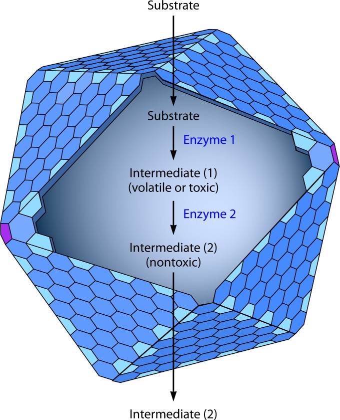
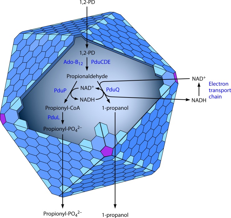
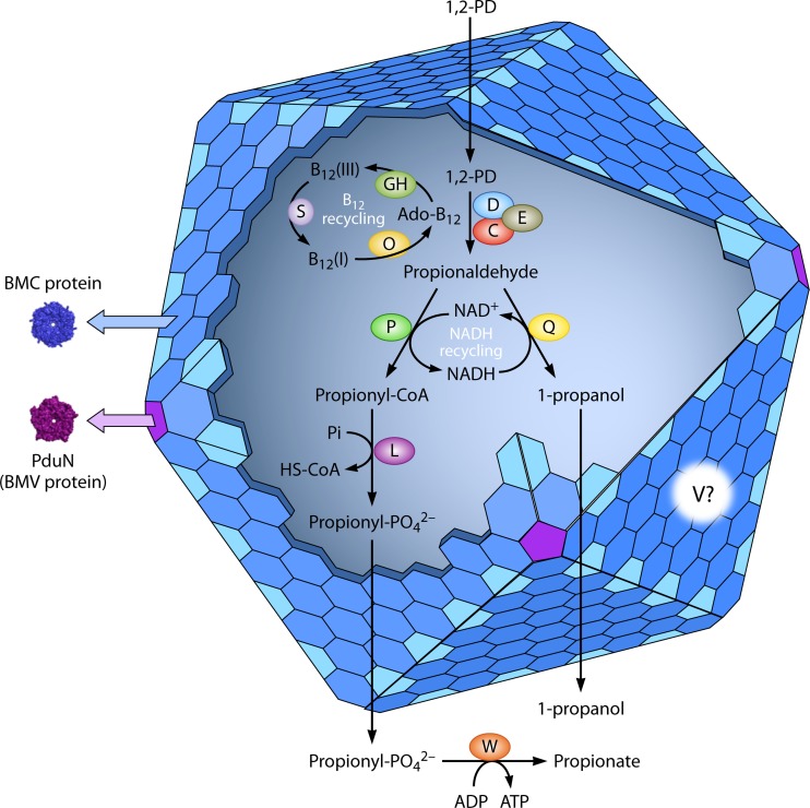
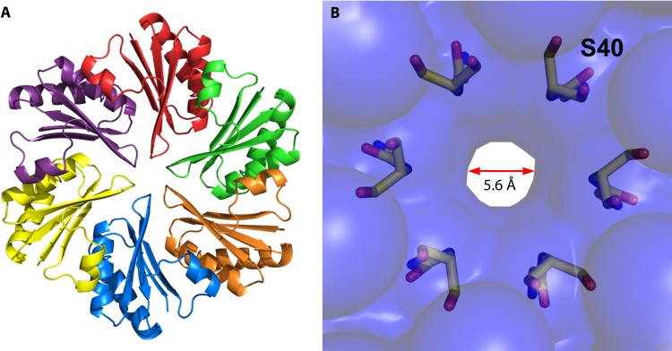
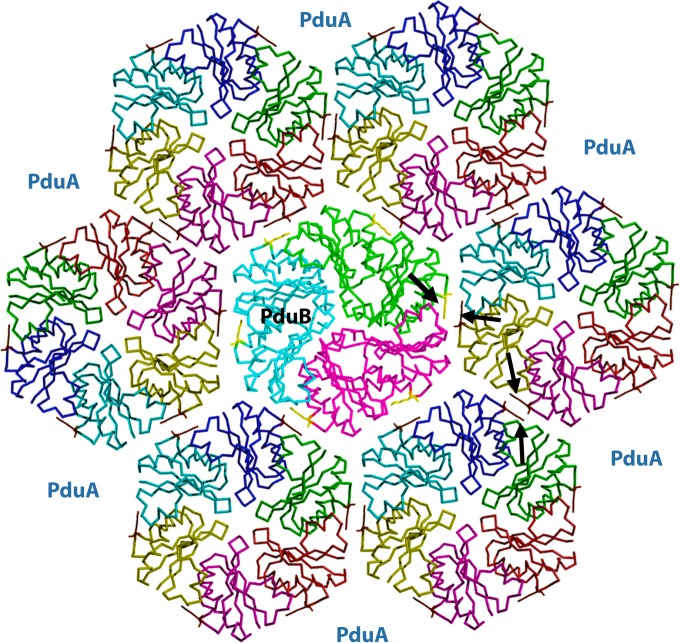
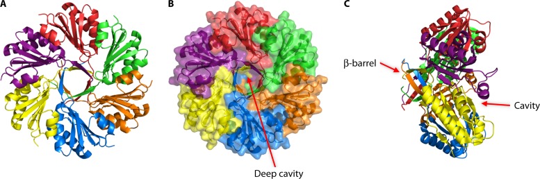
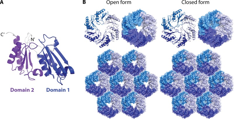
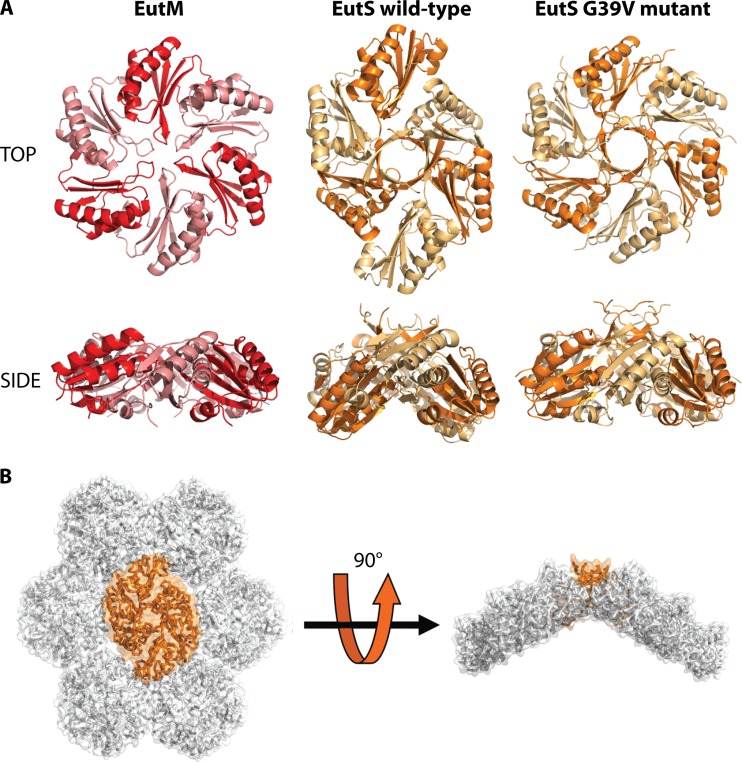
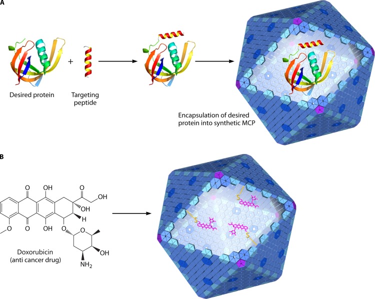
Bacterial microcompartments (MCPs) are sophisticated protein-based organelles used to optimize metabolic pathways. They consist of metabolic enzymes encapsulated within a protein shell, which creates an ideal environment for catalysis and facilitates the channeling of toxic/volatile intermediates to downstream enzymes. The metabolic processes that require MCPs are diverse and widely distributed and play important roles in global carbon fixation and bacterial pathogenesis. The protein shells of MCPs are thought to selectively control the movement of enzyme cofactors, substrates, and products (including toxic or volatile intermediates) between the MCP interior and the cytoplasm of the cell using both passive electrostatic/steric and dynamic gated mechanisms. Evidence suggests that specialized shell proteins conduct electrons between the cytoplasm and the lumen of the MCP and/or help rebuild damaged iron-sulfur centers in the encapsulated enzymes. The MCP shell is elaborated through a family of small proteins whose structural core is known as a bacterial microcompartment (BMC) domain. BMC domain proteins oligomerize into flat, hexagonally shaped tiles, which assemble into extended protein sheets that form the facets of the shell. Shape complementarity along the edges allows different types of BMC domain proteins to form mixed sheets, while sequence variation provides functional diversification. Recent studies have also revealed targeting sequences that mediate protein encapsulation within MCPs, scaffolding proteins that organize lumen enzymes and the use of private cofactor pools (NAD/H and coenzyme A [HS-CoA]) to facilitate cofactor homeostasis. Although much remains to be learned, our growing understanding of MCPs is providing a basis for bioengineering of protein-based containers for the production of chemicals/pharmaceuticals and for use as molecular delivery vehicles.


Figures

FIG 1
FIG 1
Bacterial microcompartments are used to optimize pathways with toxic or volatile intermediates. The protein shell of the microcompartment acts as a diffusion barrier that helps channel a toxic or volatile intermediate to the next pathway enzyme. In the absence of the microcompartment, such intermediates would either accumulate in the cytoplasm, causing cellular toxicity, or diffuse across the cell envelope and into the environment, resulting in the loss of valuable carbon. The carboxysome and the Pdu and Eut microcompartments help to channel CO2, propionaldehyde, and acetaldehyde, respectively. All three of these compounds pass easily through the cell envelope, and propionaldehyde and acetaldehyde are cytotoxic. In addition, MCPs may enhance catalysis by bringing enzymes, substrates, and cofactors together at high concentrations.
FIG 2
FIG 2
The enzymes encapsulated within MCPs are supplied with cofactors by internal and external recycling. Recent studies showed that the PduQ enzyme recycles NADH to NAD+ internally within the Pdu MCP. NAD+ is required for the activity of the PduP enzyme, which is essential for growth on 1,2-propanediol. However, because a PduQ mutant still grows at a rate about 50% of that of the wild type on 1,2-propanediol, external recycling is also indicated (79). In addition, studies of the Eut MCP have shown internal recycling of coenzyme A (81).
FIG 3
FIG 3
Pathway of 1,2-propanediol degradation. Under fermentative conditions, the pathway of 1,2-propanediol degradation provides a source of ATP and reducing power (NADH) but no source of cell carbon. Under aerobic conditions or during anaerobic respiration with tetrathionate, propionyl-CoA feeds into the methyl citrate pathway to provide additional ATP as well as biosynthetic building blocks.
FIG 4
FIG 4
The pdu locus of Salmonella enterica. The genes involved in 1,2-PD degradation, including those for MCP formation, cluster at a single locus. The genes for MCP formation are interspersed with those encoding pathway enzymes. This general arrangement is found for many MCP operons, although in some cases, the genes may be dispersed.
FIG 5
FIG 5
Model for 1,2-propanediol degradation by S. enterica. 1,2-PD degradation occurs within the Pdu MCP. The function of the MCP is to sequester propionaldehyde to prevent toxicity and carbon loss. The first step of 1,2-PD degradation is its conversion to propionaldehyde. The protein shell of the Pdu MCP acts as a diffusion barrier and helps to channel propionaldehyde to propionaldehyde dehydrogenase and 1-propanol dehydrogenase. It is thought that propionyl-phosphate leaves the MCP and feeds into the methyl citrate pathway, which provides additional energy and biosynthetic building blocks.
FIG 6
FIG 6
Electron micrographs of Pdu microcompartments from S. enterica. (Left) Thin sections of whole cells. (Right) Purified microcompartments.
FIG 7
FIG 7
Vitamin B12 assimilation and recycling. The first step of 1,2-propanediol degradation is catalyzed by coenzyme B12-dependent diol dehydratase. To supply diol dehydratase with its required cofactor, vitamin B12 (CN-B12) can be taken up from the environment and converted to the active coenzyme Ado-B12. During substrate turnover, catalytic by-reactions sometimes convert Ado-B12 to HO-B12, which remains enzyme bound. HO-B12 is removed from diol dehydratase by the PduGH reactivase and then converted back to Ado-B12 by a pathway similar to that used for assimilation of coenzyme B12 from the environment.
FIG 8
FIG 8
Structure of the PduO adenosyltransferase of Lactobacillus reuteri. The structure shown corresponds to the N-terminal ATR domain of PduO from Salmonella. (A) PduO in complex with cobalamin and ATP. (B) Residues interacting with ATP and cobalamin are labeled.
FIG 9
FIG 9
Structure of the PduA shell protein. (A) Hexameric structure of PduA. (B) Surface structure of PduA with pore-lining residues highlighted.
FIG 10
FIG 10
Electron micrographs of mutants impaired for formation of the Pdu microcompartment. The arrows mark normal microcompartments in wild-type S. enterica, polar bodies in the ΔpduBB′ mutant, and aggregated microcompartments in the ΔpduK mutant. (Reprinted from reference .)
FIG 11
FIG 11
Structure of PduB from Lactobacillus reuteri. Modeling indicates that coassembly of PduB and PduA mosaics helps promote stable edge interactions. In this model, a PduB trimer (center) is surrounded by six PduA hexamers. This arrangement brings lysine residues at the edges into register for favorable contacts (arrows).
FIG 12
FIG 12
The PduU shell protein is a circularly permutated hexamer whose central pore is capped by a β-barrel. (A) Ribbon model of PduU; (B) surface structure showing a deep cavity on one face of PduU; (C) side view showing a β-barrel that caps the central pore.
FIG 13
FIG 13
Comparison of wild-type microcompartments of S. enterica to those of a ΔpduM mutant. (A) Wild-type S. enterica; (B) ΔpduM deletion mutants. (Reprinted from reference .)
FIG 14
FIG 14
Pathway of coenzyme B12-dependent ethanolamine utilization. Ethanolamine degradation proceeds by a pathway analogous to that of 1,2-PD degradation (Fig. 3), the main difference being C2 intermediates rather than C3. As is the case for 1,2-PD degradation, the fermentation of ethanolamine provides a source of ATP and reducing power (NADH) but no source of cell carbon. Under aerobic conditions or during anaerobic respiration with tetrathionate, acetyl-CoA feeds into the TCA cycle to provide additional ATP and biosynthetic building blocks. *, the acetate kinase used for ethanolamine degradation is a housekeeping enzyme encoded outside the eut operon.
FIG 15
FIG 15
The ethanolamine utilization (eut) operon of S. enterica. The genes for ethanolamine degradation are found at a single locus that encodes both pathway enzymes and the proteins used to build the Eut MCP.
FIG 16
FIG 16
Hexameric structure of EutM. EutM is a good candidate for a major shell protein of the Eut MCP. Its central pore is occupied by a sulfate ion, consistent with a role as a molecular conduit for small molecules. (Reprinted from reference with permission from AAAS.)
FIG 17
FIG 17
The structure of the EutL shell protein suggests a gated pore to control the movement of metabolites from the cytoplasm to the MCP lumen. (A) EutL monomer in its closed form. (B) Open and closed configurations of EutL trimers in both ribbon diagram and surface representations. EutL forms extended protein sheets, as do a number of other MCP shell proteins (bottom). (Reprinted from reference with permission from AAAS.)
FIG 18
FIG 18
Comparison of the structures of the EutS and EutM shell protein hexamers. (A) EutM, wild-type EutS, and the EutS G39V mutant shown in two views. The wild-type EutS hexamer is bent away from a flat configuration by approximately 40°. The EutS G39V mutant is flat. (B) Hypothetical model showing how EutS (orange) might introduce curvature in an otherwise flat hexameric sheet of shell protein hexamers. (Reprinted from reference with permission from AAAS.)
FIG 19
FIG 19
Possible biotechnology applications of bacterial microcompartments. (A) Directing a protein of interest into a synthetic microcompartment might allow engineering of nanobioreactors for chemical production. (B) Bacterial microcompartments as drug delivery vehicles. Doxorubicin, an anticancer drug, might be encapsulated in the lumen of the synthetic microcompartment and targeted to diseased cells.

References

    1. Drews G, Niklowitz W. 1956. Beiträge zur Cytologie der Blaualgen. II. Zentroplasma und granulare Einschlüsse von Phormidium uncinatum. Arch. Mikrobiol. 24:147–162 - PubMed
    1. Shively JM, Ball F, Brown DH, Saunders RE. 1973. Functional organelles in prokaryotes: polyhedral inclusions (carboxysomes) of Thiobacillus neapolitanus. Science 182:584–586. 10.1126/science.182.4112.584 - DOI - PubMed
    1. Cannon GC, Shively JM. 1983. Characterization of a homogenous preparation of carboxysomes from Thiobacillus neapolitanus. Arch. Microbiol. 134:52–59. 10.1007/BF00429407 - DOI
    1. Chen P, Andersson DI, Roth JR. 1994. The control region of the pdu/cob regulon in Salmonella typhimurium. J. Bacteriol. 176:5474–5482 - PMC - PubMed
    1. Stojiljkovic I, Baeumler AJ, Heffron F. 1995. Ethanolamine utilization in Salmonella typhimurium: nucleotide sequence, protein expression, and mutational analysis of the cchA cchB eutE eutJ eutG eutH gene cluster. J. Bacteriol. 177:1357–1366 - PMC - PubMed

Publication types

Substances