. 2014 Sep 3;9(9):e106434.
doi: 10.1371/journal.pone.0106434. eCollection 2014.

Search for microRNAs expressed by intracellular bacterial pathogens in infected mammalian cells

Affiliations

Search for microRNAs expressed by intracellular bacterial pathogens in infected mammalian cells

Yuki Furuse et al. PLoS One. .

Abstract

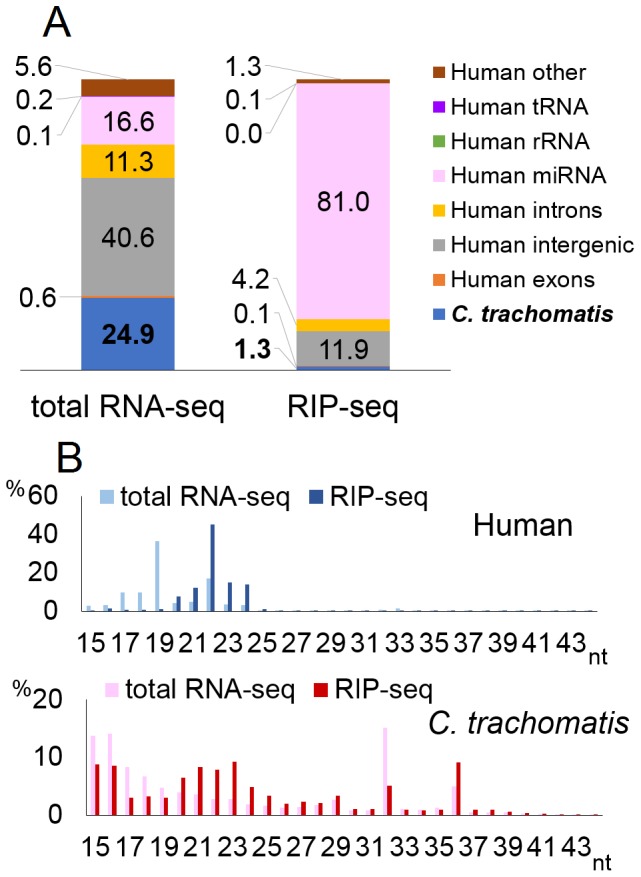
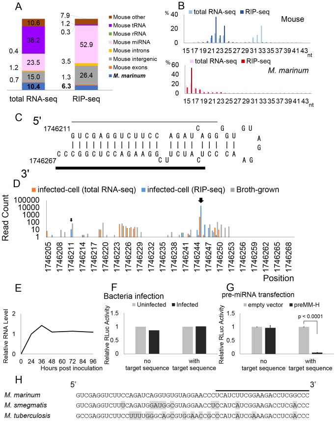
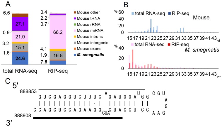
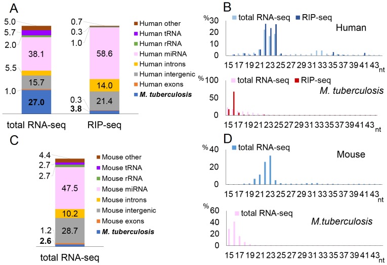
MicroRNAs are expressed by all multicellular organisms and play a critical role as post-transcriptional regulators of gene expression. Moreover, different microRNA species are known to influence the progression of a range of different diseases, including cancer and microbial infections. A number of different human viruses also encode microRNAs that can attenuate cellular innate immune responses and promote viral replication, and a fungal pathogen that infects plants has recently been shown to express microRNAs in infected cells that repress host cell immune responses and promote fungal pathogenesis. Here, we have used deep sequencing of total expressed small RNAs, as well as small RNAs associated with the cellular RNA-induced silencing complex RISC, to search for microRNAs that are potentially expressed by intracellular bacterial pathogens and translocated into infected animal cells. In the case of Legionella and Chlamydia and the two mycobacterial species M. smegmatis and M. tuberculosis, we failed to detect any bacterial small RNAs that had the characteristics expected for authentic microRNAs, although large numbers of small RNAs of bacterial origin could be recovered. However, a third mycobacterial species, M. marinum, did express an ∼ 23-nt small RNA that was bound by RISC and derived from an RNA stem-loop with the characteristics expected for a pre-microRNA. While intracellular expression of this candidate bacterial microRNA was too low to effectively repress target mRNA species in infected cultured cells in vitro, artificial overexpression of this potential bacterial pre-microRNA did result in the efficient repression of a target mRNA. This bacterial small RNA therefore represents the first candidate microRNA of bacterial origin.


Conflict of interest statement

Competing Interests: Please note that Jörn Coers & David M. Tobin are PLOS ONE Editorial Board members. This does not alter the authors' adherence to PLOS ONE Editorial policies and criteria.

Figures

Figure 1
Figure 1. Deep-sequencing of small RNAs in C. trachomatis-infected cells.
Results of deep-sequencing for C. trachomatis-infected HeLa cells. A) Gene annotation based on sequencing reads aligned to either human or bacterial genome. Values indicate the percentage of reads of each gene type in either the total or RISC IP from small RNA libraries. B) Length distribution of reads. The X-axis shows the length of small RNAs (nucleotide, nt) and the Y-axis shows the percentage of reads of each length in the total or RISC IP library.
Figure 2
Figure 2. Deep-sequencing of small RNAs in L. pneumophila-infected cells.
Results of deep-sequencing for L. pneumophila. The figures were generated as described in Fig. 1.
Figure 3
Figure 3. Deep-sequencing of small RNAs in M. marinum infected cells and characteristics of the MM-H miRNA candidate.
A and B) Results of deep-sequencing for M. marinum. The figures were generated as described in Fig. 1. C) Predicted RNA secondary structure of a possible MM-H precursor, including flanking sequences. The bold line indicates the putative mature miRNA and the thin line indicates a possible passenger strand. D) 5′ end starting positions of small RNAs from the structure shown in panel a, found in infected cell lines (total RNA-seq and RIP-seq) and in broth-grown bacteria. The Y-axis shows the absolute read count of each small RNA. Large and small arrows indicate the starting positions of the putative mature miRNA and possible passenger strand shown in panel A. E) Time-course analysis of the expression level of the MM-H RNA measured by qRT-PCR. Relative expression levels relative to the 24-h time point were normalized to the host cell U6 RNA level are indicated. Data shown represent the average of two experiments. F) Inhibitory activity of the putative M. marinum MM-H miRNA. An RLuc-based indicator, with or without two copies of a perfectly complementary target sequence for the putative MM-H miRNA inserted into the 3′ UTR, was transduced into RAW264.7 cells infected or uninfected with M. marinum. Relative RLuc activity was measured at 24 h post-transduction and then normalized to uninfected control and to the FLuc internal control, which is present in a second lentiviral vector transduced simultaneously. A representative experiment is shown. G) Similar to panel F, except in this case a Pol III-based expression vector encoding the putative MM-H pre-miRNA shown in panel C was co-transfected into 293T cells along with an RLuc-based MM-H indicator plasmid and an FLuc-based internal control. Average of three experiments with SD indicated. H) Sequence homology of the MM-H region in various mycobacteria. The predominant M. marinum MM-H sequence is indicated by a bold line. Nucleotides that differ from M. marinum are highlighted.
Figure 4
Figure 4. Deep-sequencing of small RNAs in M. smegmatis-infected cells.
A and B) Results of small RNA deep-sequencing for M. smegmatis-infected RAW264.7 cells. These figures were generated as described in Fig. 1. C) Predicted RNA secondary structure of the MM-H homology region of M. smegmatis. Bold line indicates the putative mature miRNA and the thin line a possible passenger strand found by deep-sequencing.
Figure 5
Figure 5. Deep-sequencing of small RNAs in M. tuberculosis infected cells and mice.
These figures were generated as described in Fig. 1, for in vitro infection of THP-1 cells (A and B) and in vivo infection of mice (C and D).

References

    1. Fire A, Xu S, Montgomery MK, Kostas SA, Driver SE, et al. (1998) Potent and specific genetic interference by double-stranded RNA in Caenorhabditis elegans. Nature 391: 806–811. - PubMed
    1. Elbashir SM, Lendeckel W, Tuschl T (2001) RNA interference is mediated by 21- and 22-nucleotide RNAs. Genes Dev 15: 188–200. - PMC - PubMed
    1. Martinez J, Patkaniowska A, Urlaub H, Luhrmann R, Tuschl T (2002) Single-stranded antisense siRNAs guide target RNA cleavage in RNAi. Cell 110: 563–574. - PubMed
    1. Bernstein E, Caudy AA, Hammond SM, Hannon GJ (2001) Role for a bidentate ribonuclease in the initiation step of RNA interference. Nature 409: 363–366. - PubMed
    1. Hammond SM, Bernstein E, Beach D, Hannon GJ (2000) An RNA-directed nuclease mediates post-transcriptional gene silencing in Drosophila cells. Nature 404: 293–296. - PubMed

Publication types

MeSH terms

Associated data