. 2014 Oct 7;53(39):6211-9.
doi: 10.1021/bi500850j. Epub 2014 Sep 19.

Structure and protein-protein interactions of methanol dehydrogenase from Methylococcus capsulatus (Bath)

Affiliations

Structure and protein-protein interactions of methanol dehydrogenase from Methylococcus capsulatus (Bath)

Megen A Culpepper et al. Biochemistry. .

Abstract

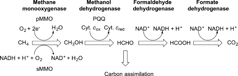
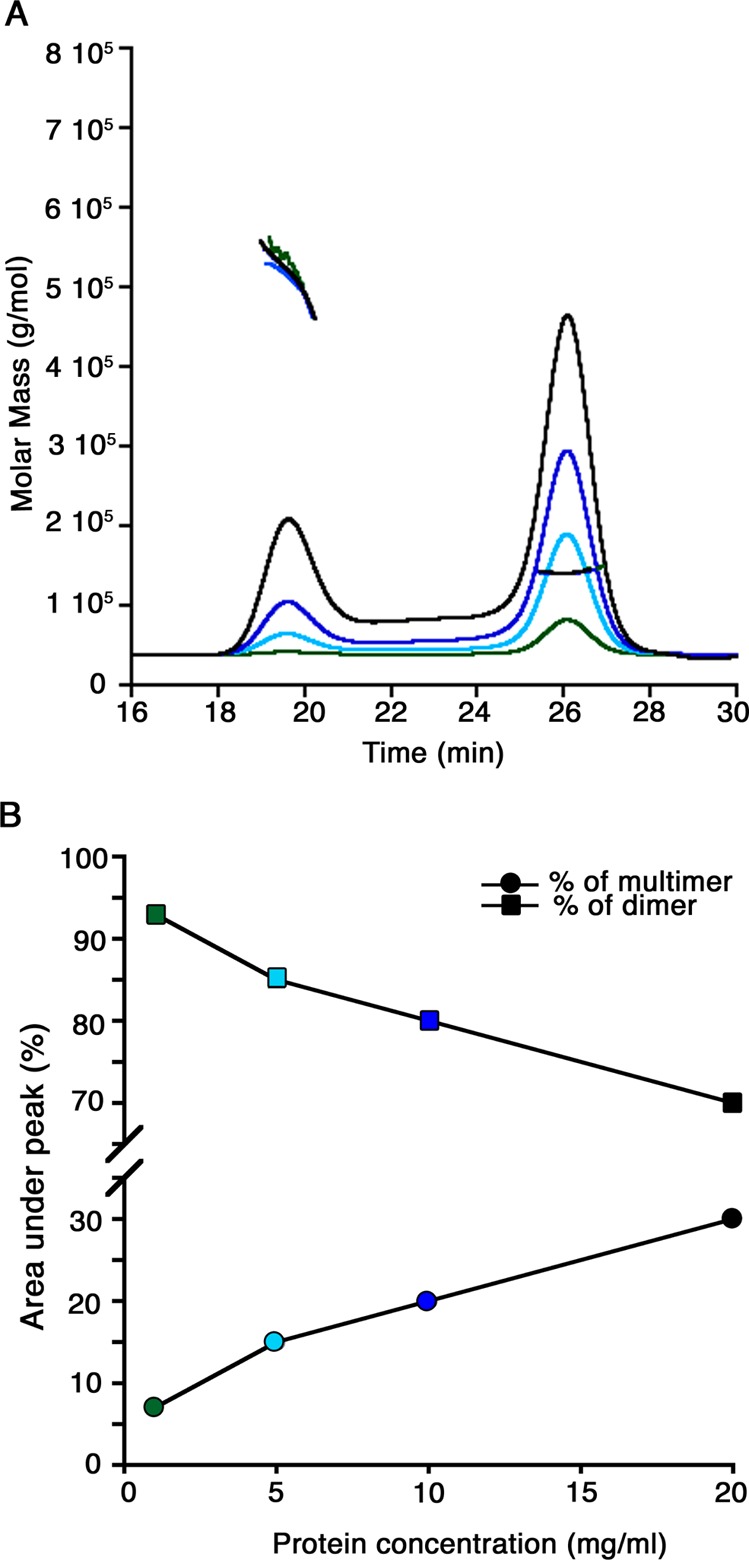
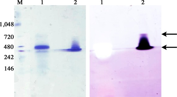
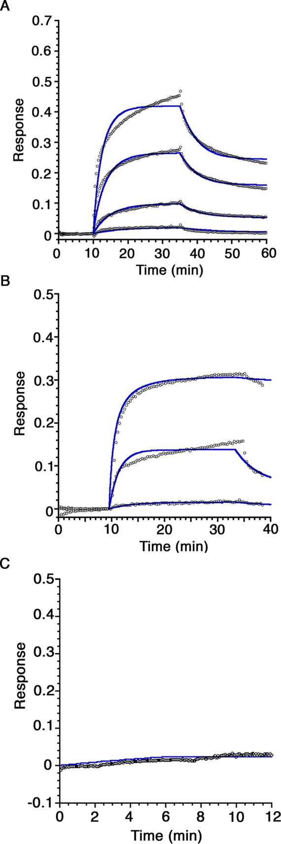
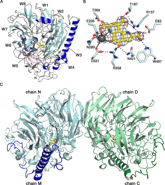
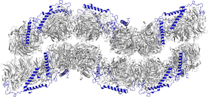
In the initial steps of their metabolic pathway, methanotrophic bacteria oxidize methane to methanol with methane monooxygenases (MMOs) and methanol to formaldehyde with methanol dehydrogenases (MDHs). Several lines of evidence suggest that the membrane-bound or particulate MMO (pMMO) and MDH interact to form a metabolic supercomplex. To further investigate the possible existence of such a supercomplex, native MDH from Methylococcus capsulatus (Bath) has been purified and characterized by size exclusion chromatography with multi-angle light scattering and X-ray crystallography. M. capsulatus (Bath) MDH is primarily a dimer in solution, although an oligomeric species with a molecular mass of ∼450-560 kDa forms at higher protein concentrations. The 2.57 Å resolution crystal structure reveals an overall fold and α2β2 dimeric architecture similar to those of other MDH structures. In addition, biolayer interferometry studies demonstrate specific protein-protein interactions between MDH and M. capsulatus (Bath) pMMO as well as between MDH and the truncated recombinant periplasmic domains of M. capsulatus (Bath) pMMO (spmoB). These interactions exhibit KD values of 833 ± 409 nM and 9.0 ± 7.7 μM, respectively. The biochemical data combined with analysis of the crystal lattice interactions observed in the MDH structure suggest a model in which MDH and pMMO associate not as a discrete, stoichiometric complex but as a larger assembly scaffolded by the intracytoplasmic membranes.


Figures

Figure 1
Figure 1
Metabolic pathway of M. capsulatus (Bath).
Figure 2
Figure 2
Oligomerization states of M. capsulatus (Bath) MDH in solution. (A) SEC-MALS analysis of MDH at varying protein concentrations. The signals from the refractive index detector are shown as a function of elution time (black for 20 mg/mL, blue for 10 mg/mL, cyan for 5 mg/mL, and green for 1 mg/mL). The thick horizontal lines indicate the calculated molecular masses of the eluting peaks. (B) Comparison of the multimer and dimer peak areas (as percentages) from the SEC-MALS experiments displayed as a line graph. An increased protein concentration results in an increase in multimer percentage concomitant with a decrease in dimer percentage. The colors of the data points correspond to the SEC-MALS traces in panel A.
Figure 3
Figure 3
D-PAGE analysis of pMMO and MDH from M. capsulatus (Bath). Gels were either stained with Coomassie dye (left) or reacted with PMS, NBT, and methanol to detect MDH activity (right). Equal amounts (25 μg) of pMMO (lane 1) and MDH (lane 2) were loaded per gel. Molecular mass markers are shown in lane M (kDa). The arrows indicate the putative MDH dimer and oligomer species.
Figure 4
Figure 4
Biolayer interferometry sensorgrams of the protein–protein interactions of immobilized M. capsulatus (Bath) MDH. (A) Purified M. capsulatus (Bath) pMMO samples at concentrations of 0.5, 1, 2.5, and 5 μM were monitored for binding to immobilized MDH. The data fitting curves are displayed as blue lines. (B) Refolded spmoB samples at concentrations of 0.3, 3, and 30 μM were reacted with immobilized MDH. (C) Immobilized BSA was monitored for interaction with 30 μM spmoB. No binding response is observed. Each experiment was repeated with three independent biological samples.
Figure 5
Figure 5
Structure of M. capsulatus (Bath) MDH. (A) αβ protomer. The eight-bladed β sheet propeller motif is colored light blue, and the blades are labeled W1–W8. Auxiliary β strands are colored light pink and α helices blue, including the long α helix of the β subunit. A PQQ ligand (yellow) and Ca2+ ion (gray) are located in the cavity of the propeller core fold. (B) Composite omit map generated in Phenix contoured at 1σ showing the cofactors and hydrogen bonding residues. Ligand coordination to the Ca2+ ion is shown as solid lines, and hydrogen bonds are shown as dashed lines. (C) α2β2 dimer structure with chains C and D colored light and dark green and chains M and N colored light and dark blue.
Figure 6
Figure 6
Crystal packing interactions of MDH from M. capsulatus (Bath). Symmetry-related molecules of MDH reveal a bilayer-like packing arrangement with the β subunit exposed to the solvent. The α subunit is colored gray and the β subunit blue.

References

    1. Hanson R. S.; Hanson T. E. (1996) Methanotrophic bacteria. Microbiol. Rev. 60, 439–471. - PMC - PubMed
    1. Semrau J. D.; Dispirito A. A.; Yoon S. (2010) Methanotrophs and copper. FEMS Microbiol. Lett. 34, 496–531. - PubMed
    1. Fei Q.; Guarnieri M. T.; Tao L.; Laurens L. M.; Dowe N.; Pienkos P. T. (2014) Bioconversion of natural gas to liquid fuel: Opportunities and challenges. Biotechnol. Adv. 32, 596–614. - PubMed
    1. Hakemian A. S.; Rosenzweig A. C. (2007) The biochemistry of methane oxidation. Annu. Rev. Biochem. 76, 223–241. - PubMed
    1. Culpepper M. A.; Rosenzweig A. C. (2012) Architecture and active site of particulate methane monooxygenase. Crit. Rev. Biochem. Mol. Biol. 47, 483–492. - PMC - PubMed

Publication types

MeSH terms