. 2014 Sep 2;107(5):1129-1135.
doi: 10.1016/j.bpj.2014.07.016.

Surfactant bilayers maintain transmembrane protein activity

Affiliations

Surfactant bilayers maintain transmembrane protein activity

Gamal Rayan et al. Biophys J. .

Abstract

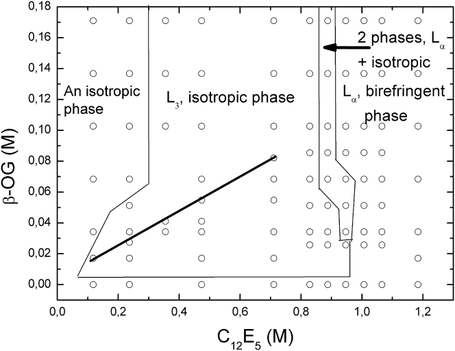
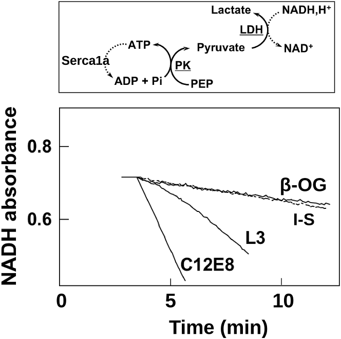
In vitro studies of membrane proteins are of interest only if their structure and function are significantly preserved. One approach is to insert them into the lipid bilayers of highly viscous cubic phases rendering the insertion and manipulation of proteins difficult. Less viscous lipid sponge phases are sometimes used, but their relatively narrow domain of existence can be easily disrupted by protein insertion. We present here a sponge phase consisting of nonionic surfactant bilayers. Its extended domain of existence and its low viscosity allow easy insertion and manipulation of membrane proteins. We show for the first time, to our knowledge, that transmembrane proteins, such as bacteriorhodopsin, sarcoplasmic reticulum Ca(2+)ATPase (SERCA1a), and its associated enzymes, are fully active in a surfactant phase.


Figures

Figure 1
Figure 1
Partial phase diagrams of the ternary systems consisting of C12E5, water, and β-OG as cosurfactant. Each symbol corresponds to an experimental sample that was investigated at 20°C. The lines drawn on the diagram represent approximate boundaries between the phases. The black line in the L3 region of the diagram corresponds to the dilution line that was investigated with the following methods; small angle x-ray scattering, small angle neutron scattering, FFEM, and FRAPP. Throughout this black line the L3 phase exists from 6 to 30°C.
Figure 2
Figure 2
Typical decrease of the FRAPP fringes contrast as a function of time (here for SERCA1a in L3). From the fit of this curve (measured at an interfringe distance i = 19.4 μm) we obtain a characteristic decay time τ = 5.2 s, which corresponds to a D value of 1.7 ± 0.1 μm/s2. In the inset, we present the variation of 1/τ as a function of (i/2π)2. The linear behavior indicates the Brownian motion of the protein embedded in the bilayers of L3 phase and the slope of the linear fit leads to the self-diffusion coefficient of the protein.
Figure 3
Figure 3
Freeze fracture electron microscopy of the L3 containing (a) SERCA1a at 2.5 μM and (b) BR at 5.0 μM. The image represents the midplane of the bilayers of the sponge phase and the spots pointed by the arrows represent the replica of SERCA and BR proteins embedded in the bilayer. The stars point out to the region of the phase devoid of proteins.
Figure 4
Figure 4
Time-resolved absorption of BR at 415 nm dissolved in the purple membrane (▴); β-OG micelles (☆); the L3 phase (CBR =2.1 μM) (▪); the solution mimicking the intermembrane liquid space (CBR = 0.1 μM) (□), see Results and Discussion.
Figure 5
Figure 5
Enzymatic activity of the SERCA1a reconstituted in various environments. The end product of this coupled-enzyme assay is the oxidation of NADH to NAD+, which is seen as the decrease in absorption at 340 nm (the top panel). In the bottom panel the activity of the enzyme in the L3 phase is compared to its activity in micellar solutions of either β-OG (low activity), C12E8 (high activity), or in a medium mimicking the L3 intermembrane space (I-S, dotted line, low activity). The latter measurement has been performed at an enzyme concentration of 0.1 μM corresponding to the threshold detection by FRAPP. All other measurements have been performed at 1 μM. ATP: adenosine triphosphate. ADP: adenosine diphosphate. NAD: nicotinamide adenine dinucleotide. PK: pyruvate kinase. PEP: phosphoenolpyruvic acid.

References

    1. Caffrey M. Crystallizing membrane proteins for structure determination: use of lipidic mesophases. Annu. Rev. Biophys. 2009;38:29–51. - PubMed
    1. Caffrey M. Crystallizing membrane proteins for structure-function studies using lipidic mesophases. Biochem. Soc. Trans. 2011;39:725–732. - PMC - PubMed
    1. Hyde S.T. Identification of lyotropic liquid crystalline mesophases. In: Holmberg K., editor. Handbook of Applied Surface and Colloid Chemistry. John Wiley & Sons; West Sussex: 2001. pp. 299–332.
    1. Caffrey M. Membrane protein crystallization in lipidic bicontinuous liquid crystals. In: Lynch M.L., Spicer P.T., editors. Bicontinuous Liquid Crystals. CRC Press; Boca Raton, FL: 2005. pp. 307–319.
    1. Caffrey M., Cherezov V. Crystallizing membrane proteins using lipidic mesophases. Nat. Protoc. 2009;4:706–731. - PMC - PubMed

Publication types

LinkOut - more resources