

The mechanism of collapse of heterogeneous lipid monolayers
- PMID: 25185549
- PMCID: PMC4156665
- DOI: 10.1016/j.bpj.2014.05.053
The mechanism of collapse of heterogeneous lipid monolayers
Abstract
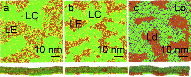
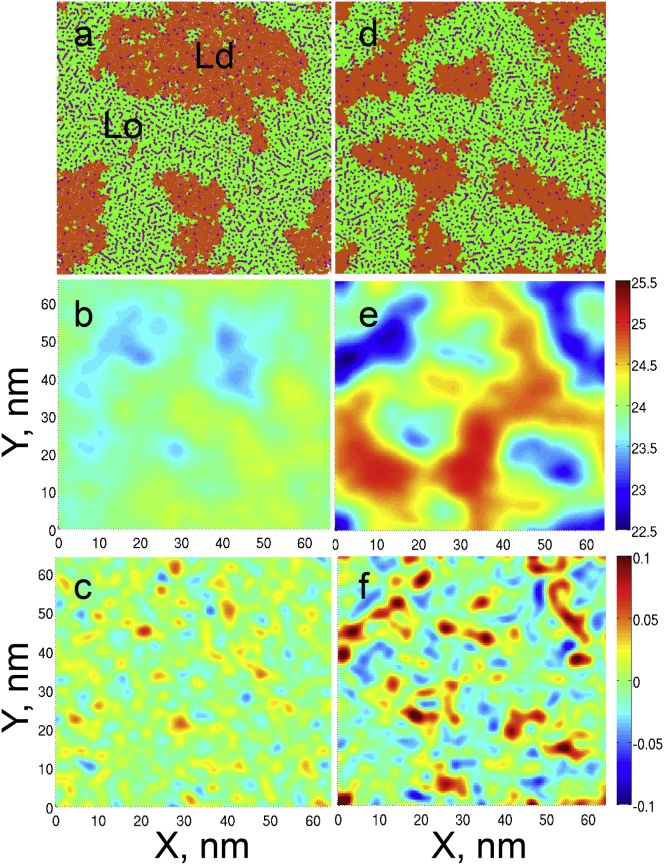
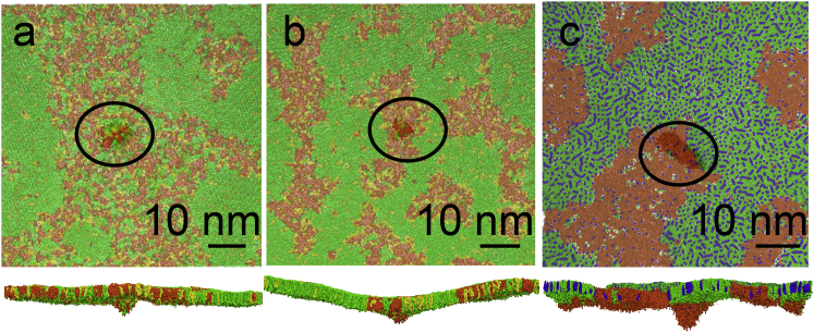
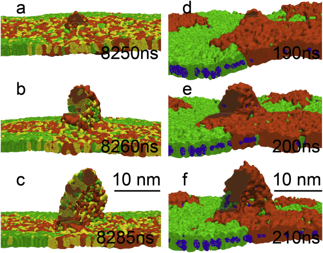
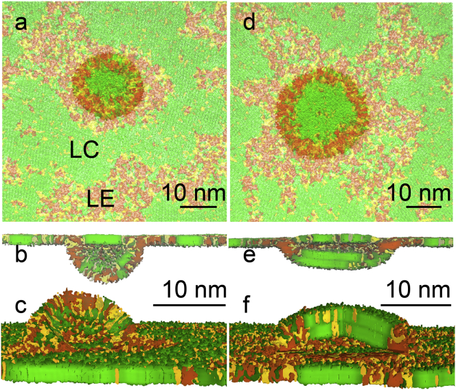
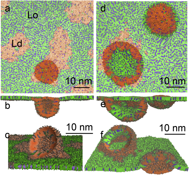
Collapse of homogeneous lipid monolayers is known to proceed via wrinkling/buckling, followed by folding into bilayers in water. For heterogeneous monolayers with phase coexistence, the mechanism of collapse remains unclear. Here, we investigated collapse of lipid monolayers with coexisting liquid-liquid and liquid-solid domains using molecular dynamics simulations. The MARTINI coarse-grained model was employed to simulate monolayers of ∼80 nm in lateral dimension for 10-25 μs. The monolayer minimum surface tension decreased in the presence of solid domains, especially if they percolated. Liquid-ordered domains facilitated monolayer collapse due to the spontaneous curvature induced at a high cholesterol concentration. Upon collapse, bilayer folds formed in the liquid (disordered) phase; curved domains shifted the nucleation sites toward the phase boundary. The liquid (disordered) phase was preferentially transferred into bilayers, in agreement with the squeeze-out hypothesis. As a result, the composition and phase distribution were altered in the monolayer in equilibrium with bilayers compared to a flat monolayer at the same surface tension. The composition and phase behavior of the bilayers depended on the degree of monolayer compression. The monolayer-bilayer connection region was enriched in unsaturated lipids. Percolation of solid domains slowed down monolayer collapse by several orders of magnitude. These results are important for understanding the mechanism of two-to-three-dimensional transformations in heterogeneous thin films and the role of lateral organization in biological membranes. The study is directly relevant for the function of lung surfactant, and can explain the role of nanodomains in its surface activity and inhibition by an increased cholesterol concentration.
Copyright © 2014 Biophysical Society. Published by Elsevier Inc. All rights reserved.
Figures
Comment in
-
New insights into lipid monolayers from coarse-grained simulation techniques.Biophys J. 2014 Sep 2;107(5):1038-1039. doi: 10.1016/j.bpj.2014.07.030. Biophys J. 2014. PMID: 25185538 Free PMC article. No abstract available.
References
-
- Tanford C. Wiley; New York: 1980. The Hydrophobic Effect.
-
- Mouritsen O.G. Springer; Heidelberg, Germany: 2005. Life—As a Matter of Fat.
-
- Knobler C.M., Desai R.C. Phase transitions in monolayers. Annu. Rev. Phys. Chem. 1992;43:207–236.
-
- McConnell H.M. Structures and transitions in lipid monolayers at the air-water-interface. Annu. Rev. Phys. Chem. 1991;42:171–195.
-
- Debrégeas G., de Gennes P.-G., Brochard-Wyart F. The life and death of “bare” viscous bubbles. Science. 1998;279:1704–1707. - PubMed
Publication types
MeSH terms
Substances
LinkOut - more resources
Full Text Sources
Other Literature Sources
Research Materials
