. 2014 Sep 4:5:4807.
doi: 10.1038/ncomms5807.

Dynactin functions as both a dynamic tether and brake during dynein-driven motility

Affiliations

Dynactin functions as both a dynamic tether and brake during dynein-driven motility

Swathi Ayloo et al. Nat Commun. .

Abstract

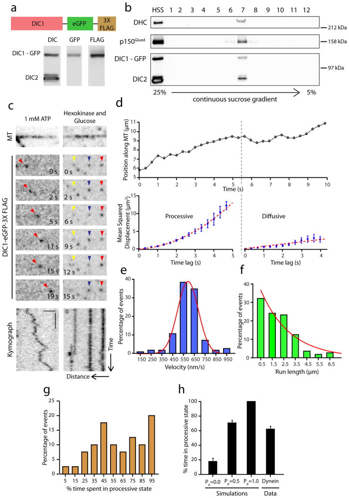
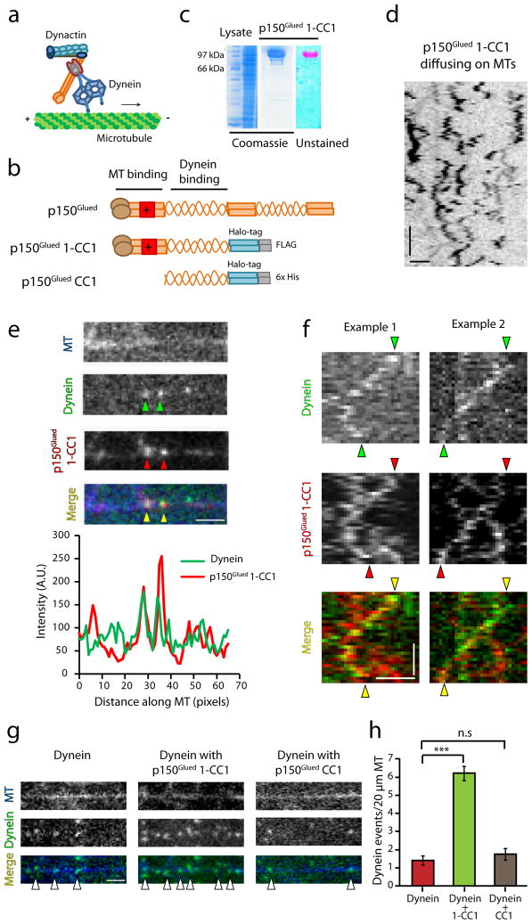
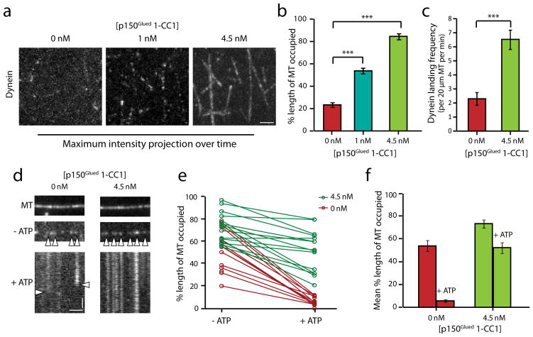
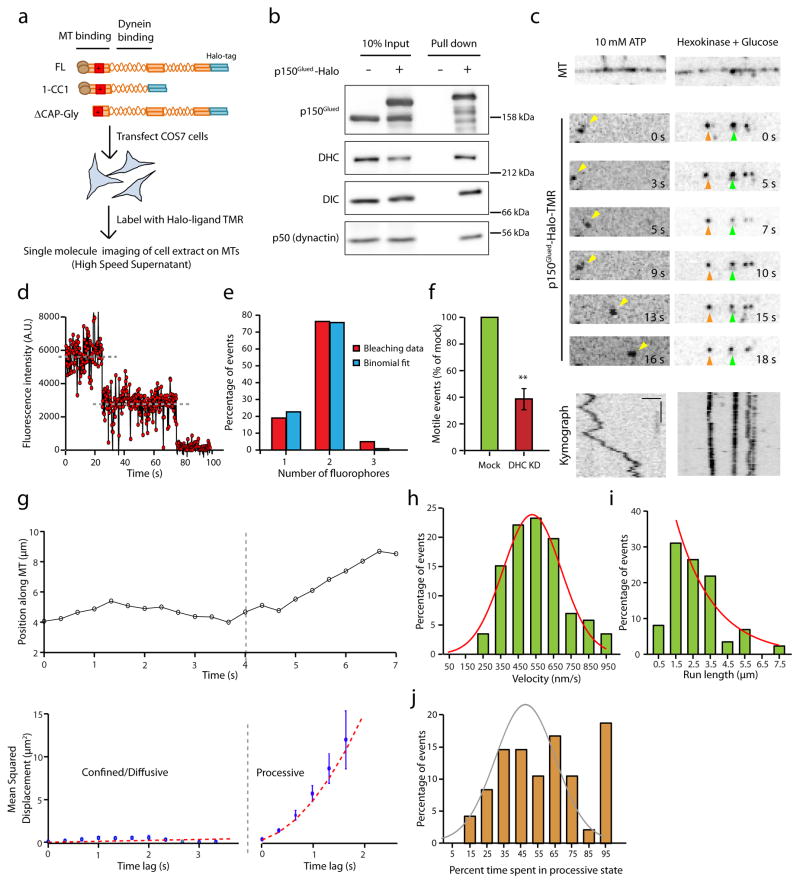
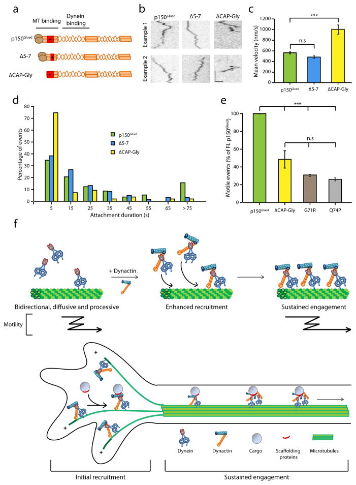
Dynactin is an essential cofactor for most cellular functions of the microtubule motor cytoplasmic dynein, but the mechanism by which dynactin activates dynein remains unclear. Here we use single molecule approaches to investigate dynein regulation by the dynactin subunit p150(Glued). We investigate the formation and motility of a dynein-p150(Glued) co-complex using dual-colour total internal reflection fluorescence microscopy. p150(Glued) recruits and tethers dynein to the microtubule in a concentration-dependent manner. Single molecule imaging of motility in cell extracts demonstrates that the CAP-Gly domain of p150(Glued) decreases the detachment rate of the dynein-dynactin complex from the microtubule and also acts as a brake to slow the dynein motor. Consistent with this important role, two neurodegenerative disease-causing mutations in the CAP-Gly domain abrogate these functions in our assays. Together, these observations support a model in which dynactin enhances the initial recruitment of dynein onto microtubules and promotes the sustained engagement of dynein with its cytoskeletal track.


Conflict of interest statement

Competing financial interests

The authors declare no competing financial interests.

Figures

Figure 1
Figure 1. Dynein-GFP switches stochastically from processive to diffusive states of motion
(a) Schematic of the DIC1-eGFP-FLAG gene knocked into the DIC1 locus. High speed supernatant (HSS) of homogenized mouse brain tissue was probed for DIC, GFP and FLAG. (b) Immunoblot analysis of sucrose gradient purified dynein. DIC1-GFP-FLAG is efficiently incorporated into the dynein complex as indicated by co-sedimentation with dynein heavy chain (DHC) at 20S. (c) Time series and corresponding kymograph showing projection of the movement of the particle over time for a single dynein-GFP molecule at 1 mM Mg-ATP and in the presence of hexokinase and glucose (ATP depletion system). Horizontal bar, 2 μm. Vertical bar, 5 s. All the images are contrast inverted to represent the signal as black on a white background. (d) A representative trajectory of a dynein-GFP molecule. Following gradient analysis for node detection (see Methods), mean squared displacement (MSD) analysis shows that the motion switches from processive (MSD = v2t2) to a diffusive state (MSD = 2Dt). Error bars indicate SEM. (e and f) Distributions of velocities and run lengths of particles tracked (n=112, 3 independent experiments). Curves represent a gaussian distribution and an exponential decay curve respectively. (g) Frequency distribution for the amount of time the particles (n=40, 2 independent experiments) spent in a processive state. On average, about 60% of the time, the motion is processive. (h) Quantitation of the percent time spent in processive state for simulated tracks alongside the average for experimental data obtained from Fig. 1g. Mean ± SEM. N=50 for the simulations and n=40 for the experimental data.
Figure 2
Figure 2. p150Glued co-migrates with dynein and enhances its recruitment onto microtubules
(a) Schematic illustrating the interaction of dynein with dynactin. (b) Schematic of the recombinant constructs p150Glued 1-CC1 and p150Glued CC1 engineered with a Halo tag and affinity tags at the C-terminus (c) Coomassie stained gel showing the lysate and the protein p150Glued 1-CC1 after the final purification step. The unstained gel shows the labeling of the protein with TMR. (d) Contrast inverted kymograph of p150Glued 1-CC1 diffusing on microtubules. Horizontal bar, 2 μm. Vertical bar, 5 s. (e) Co-localization of dynein and p150Glued 1-CC1 on microtubules and corresponding line-scan intensity profile. (f) Representative kymographs of co-migration of dynein with p150Glued 1-CC1. Horizontal bar, 2 μm. Vertical bar, 10 s. (g) Addition of p150Glued 1-CC1 increases the landing events of dynein while p150Glued CC1 has no effect on the recruitment of dynein. Horizontal bar, 2 μm. (h) Quantitation of the landing events of dynein in each condition shows that p150Glued 1-CC1 increases the landing events greater than 4-fold. Mean ± SEM, n=25, 2 independent experiments, ***p<0.001, one-way ANOVA with Tukey’s post-hoc test. Concentration of the recombinant protein used in each case is 4.5 nM.
Figure 3
Figure 3. p150Glued regulates the association and dissociation of dynein from microtubules
(a) Maximum-intensity projection over time of dynein motility (movies recorded for 2 min) in the presence of p150Glued 1-CC1. Horizontal bar, 2 μm. (b) Quantitation of the percent occupancy of microtubules using line-scan intensity analysis. Mean ± SEM, n=20, 2 independent experiments, ***p<0.001, one-way ANOVA with Tukey’s post-hoc test. (c) Addition of p150Glued 1-CC1 increases the landing frequency of dynein. Mean ± SEM, n=20, 2 independent experiments, ***p<0.001, Student’s t-test. (d) Representative images of dynein bound to microtubules in the absence of ATP and kymographs following 5 mM Mg-ATP wash. Horizontal bar, 2 μm. Vertical bar, 10 s. (e) Quantitation of the percent occupancy of microtubules before and after the ATP wash. The corresponding data points for the same microtubules before and after the ATP wash are connected by a line, n=15 for each condition, 2 independent experiments. (f) Average of the data shown in (e). The control had a 10-fold change post ATP wash while the addition of p150Glued 1-CC1 reduced the fold change to ~1.5 ***p<0.001, Student’s t-test.
Figure 4
Figure 4. The motility of p150Glued in cell extracts is dynein-driven and ATP-dependent
(a) Schematic of the experimental design with an illustration of p150Glued constructs. (b) COS7 cell extracts were precipitated with a HaloLink resin that binds specifically to the Halo tag. Protein complexes bound to the resin were analyzed by immunoblotting. The dynein complex co-precipitates with p150Glued-Halo and the addition of the Halo tag at the C-terminus does not interfere with the incorporation of p150Glued into the dynactin complex (as shown by p50). (c) Representative time series and corresponding kymographs of the motility of p150Glued-Halo in 10 mM Mg-ATP and in the presence of hexokinase and glucose (an ATP depletion system). Horizontal bar, 2 μm. Vertical bar, 5 s. All the images are contrast inverted. (d) A representative time course of photobleaching of TMR labeled p150Glued shows two decay steps. Grey lines indicate the steps. (e) Frequency distribution of the photobleaching steps for n=42 particles fit with a binomial distribution. (f) Quantitation of the number of active, motile events of p150Glued-Halo in the mock and dynein knock-down conditions. The number of events was normalized to the length of the microtubules and the expression levels of p150Glued-Halo in extracts (densitometry analysis using ImageJ). DHC knock-down resulted in a reduction in the number of motile events. Mean ± SEM, n=3 experiments, **p<0.01, Student’s t-test. (g) A representative trajectory of p150Glued-Halo with its corresponding MSD analysis. Error bars indicate SEM. (h and (i) Velocity and run length distribution of the tracked p150Glued particles. Tracks (n=60, 3 independent experiments) were parsed into processive and diffusive states of motion using GrAND and velocities were calculated for the processive segments only. Curved lines indicate Gaussian and exponential decay fit respectively. (j) Frequency distribution for the amount of time p150Glued-Halo spent in a processive state. Two populations can be observed - about 20% that are processive throughout the motion along the microtubule and the remaining that display both processive and diffusive phases. A Gaussian fit (shown in gray) excluding the last data point improves the R2 from 0.45 to 0.81 suggesting there could be two populations.
Figure 5
Figure 5. The CAP-Gly domain of dynactin enhances the engagement of dynein onto microtubules
(a) Schematic of the Δ5-7 and ΔCAP-Gly constructs of p150Glued. While the Δ5-7 lacks much of the basic domain, ΔCAP-Gly is a deletion of the CAP-Gly domain. (b) Representative kymographs of the motility of p150Glued, Δ5-7 and ΔCAP-Gly constructs in complex with dynein along microtubules. Horizontal bar, 2 μm. Vertical bar, 5 s. Kymographs are contrast inverted. (c) Mean velocities of all the particles tracked for p150Glued, Δ5-7 and ΔCAP-Gly. Mean ± SEM, n=60 for p150Glued and Δ5-7, n=40 for ΔCAP-Gly, 3 independent experiments, ***p<0.001, one-way ANOVA with Tukey’s post-hoc test. (d) Frequency distribution of the binding times of Δ5-7 and ΔCAP-Gly in comparison to p150Glued. Greater than 70% of the ΔCAP-Gly particles have a binding time of less than 10 seconds (n=60 for each, 3 independent experiments). (e) Quantitation of the number of active, motile events of the ΔCAP-Gly and two mutant G71R and Q74P in comparison to the full-length p150Glued. The number of events was normalized to the length of the microtubules and the expression levels of constructs in cell extracts (densitometry analysis using ImageJ). Mean ± SEM, n=3 independent experiments, ***p<0.001, one-way ANOVA with Tukey’s post-hoc test. (f) Model for the regulation of dynein by dynactin depicting two functions of dynactin – initial recruitment and sustained engagement of the dynein motor.

References

    1. Schroer TA. Dynactin. Annu Rev Cell Dev Biol. 2004;20:759–79. - PubMed
    1. Gill SR, et al. Dynactin, a conserved, ubiquitously expressed component of an activator of vesicle motility mediated by cytoplasmic dynein. J Cell Biol. 1991;115:1639–50. - PMC - PubMed
    1. Schafer DA, Gill SR, Cooper JA, Heuser JE, Schroer T. A Ultrastructural analysis of the dynactin complex: an actin-related protein is a component of a filament that resembles F-actin. J Cell Biol. 1994;126:403–12. - PMC - PubMed
    1. Holzbaur ELF, et al. Homology of a 150K cytoplasmic dynein- associated polypeptide with the Drosophila gene Glued. Nature. 1991;351:579–583. - PubMed
    1. Karki S, Holzbaur ELF. Affinity Chromatography Demonstrates a Direct Binding between Cytoplasmic Dynein and the Dynactin Complex. J Biol Chem. 1995;270:28806–28811. - PubMed

Publication types

MeSH terms

LinkOut - more resources