. 2014 Oct;26(10):1494-507.
doi: 10.1111/nmo.12419. Epub 2014 Sep 3.

Distribution across tissue layers of extrinsic nerves innervating the mouse colorectum - an in vitro anterograde tracing study

Affiliations

Distribution across tissue layers of extrinsic nerves innervating the mouse colorectum - an in vitro anterograde tracing study

Pablo R Brumovsky et al. Neurogastroenterol Motil. 2014 Oct.

Abstract

Background: Anterograde in vitro tracing of the pelvic nerve (PN) and visualization in the horizontal plane in whole mount preparations has been fundamental in the analysis of distribution of peripheral nerves innervating the colorectum. Here, we performed a similar analysis, but in cryostat sections of the mouse colorectum, allowing for a more direct visualization of nerve distribution in all tissue layers.

Methods: Colorectum with attached PNs was dissected from adult male BalbC mice. Presence of active afferents was certified by single fiber recording of fine PN fibers. This was followed by 'bulk' (all fibers) anterograde tracing using biotinamide (BTA). Histo- and immunohistochemical techniques were used for visualization of BTA-positive nerves, and evaluation of co-localization with calcitonin gene-related peptide (CGRP), respectively. Tissue was analyzed using confocal microscopy on transverse or longitudinal colorectum sections.

Key results: Abundant BTA-positive nerves spanning all layers of the mouse colorectum and contacting myenteric plexus neurons, distributing within the muscle layer, penetrating deeper into the organ and contacting blood vessels, submucosal plexus neurons or even penetrating the mucosa, were regularly detected. Several traced axons co-localized CGRP, supporting their afferent nature. Finally, anterograde tracing of the PN also exposed abundant BTA-positive nerves in the major pelvic ganglion.

Conclusions & inferences: We present the patterns of innervation of extrinsic axons across layers in the mouse colorectum, including the labile mucosal layer. The proposed approach could also be useful in the analysis of associations between morphology and physiology of peripheral nerves targeting the different layers of the colorectum.

Keywords: anterograde; biotinamide; colorectum; in vitro tracing; peripheral nerves; primary afferents.


Conflict of interest statement

DISCLOSURE

None of the authors have any actual or potential conflict of interest.

Figures

Figure 1
Figure 1
Tissue preparation and fibre recording. (A) Mouse pelvic cavity showing partially dissected organs during collection of tissue for in vitro PN recording and anterograde tracing. B) PN (black arrow) tributaries (white arrows) to the colorectum are dissected and shown, penetrating the organ at an angle. C) Colorectum opened and pinned flat mucosal side up in a Sylgard®-lined organ chamber perfused with 32°C Krebs solution. The paraffin-filled recording chamber is isolated from the organ chamber by a removable plastic gate with a mouse-hole through which the PN and accompanying sciatic nerve passes. A PN fine fibre is shown (arrow), previous to its attachment to the recording electrode (white double arrowhead). For illustration purposes, the location of a number of receptive fields tested during the experiment is shown by probing using a carbon-coated von Frey hair (black double arrowheads). (D) The PN after splitting into fine fibres (dotted line shows area covered by BTA). (E) Examples of electrophysiological recording from three different fine fibers (1–3), showing the response patterns to blunt stroking (using a camel’s hair brush), 400 mg probing using a von Frey filament, gross stretching, 10 mg stroking using a calibrated taklon brush, or exposure to an inflammatory soup (see [18]).
Figure 2
Figure 2
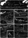
Distribution of BTA-positive nerves terminating in different layers of the mouse colorectum. Confocal fluorescence photomicrographs of longitudinal (A, C–G) or transverse (B) sections of the colorectum incubated with streptavidin, alexa fluor 488. (A) Thick and intensely stained BTA-positive nerve fibres of passage are seen in the proximity of the colorectal surface (white arrows), along with thinner traced nerves associated with the mesentery (arrowheads). A fine BTA-positive nerve fibre in the underlying myenteric plexus is also detected (black arrows). B) Abundant BTA-positive nerves are detected in the submucosal (white double arrowheads) and mucosal layers (black double arrowheads), as well as in relation to blood vessels (black arrowhead; asterisks indicate the lumen of blood vessels). (C–G) Various examples of BTA-positive nerves distributing within the myenteric plexus (black arrows in C, D, E) or traversing the circular muscle layer (double arrows in C, E, F). Note presence of thick (double arrow in E) and thin (double arrow in G; shown at higher magnification in G) traversing BTA-positive nerves changing direction in an abrupt angle, and extending for several micrometers with an orientation parallel to the myenteric plexus within the submucosal layer. Additional BTA-positive nerves innervating blood vessels (black arrowhead in E, F; asterisks show blood vessels) or traveling within the submucosal plexus underneath the mucosa (white double arrowhead in G). Scale bars: 50 µm (A–E, G); 20 µm (F, inset in G).
Figure 3
Figure 3
Identification of BTA-positive colorectal nerves in association to neurons in the myenteric and submucosal plexuses, or to blood vessels. Confocal immunofluorescence photomicrographs of longitudinal sections of the colorectum incubated with streptavidin, alexa fluor 488 (A, D, G, J, M), co-incubated with propidium iodide(B, E, H, K, N) (C, F, I, L, O show merged photomicrographs). (A–F) BTA-positive nerves (black arrows in A, D) are shown in close relationship with myenteric plexus neurons (white arrows in B, E). A nerve bundle previously associated with the myenteric plexus traverses the circular muscle layer (double arrow in D) before dividing into two fibres penetrating the submucosal layer (double arrowheads in D). (G–I; shown at higher magnification in J–L) BTA-positive nerves (black arrowheads in G, J) are seen in close association with blood vessels (arrows in H, K). (M–O) A BTA-positive nerve (double arrowheads) reaching submucosal plexus neurons is shown. Scale bars: 10 µm (C=A, B; F=D, E); 20 µm (L=J, K; O=M, N); 50 µm (I=G, H).
Figure 4
Figure 4
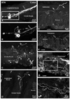
Distribution in the myenteric plexus, the circular muscle and mucosal layers, of BTA-positive nerves terminating in the mouse colorectum. Confocal fluorescence photomicrographs of longitudinal (A–C, E–H) or transverse (D) sections of the colorectum incubated with streptavidin, alexa fluor 488. (A; shown at higher magnification in B) Intensely stained BTA-positive varicose nerve endings (black arrows) are seen terminating in the myenteric plexus (black asterisks). A presumable viscerofugal neuron is also observed (white asterisk) (C) An apparently single BTA-positive axon (double arrows) penetrates and subdivides into multiple varicose nerve endings (arrowheads) within the circular muscle. (D) What appears to be a single BTA-positive axon is seen dividing into a projection towards the circular muscle layer (double arrow) and the submucosal layer (double arrowhead). (E–H) Several BTA-positive nerves, often of small calibre, are detected in the mucosal layer (black double arrowhead), often associated with traced nerves in the submucosal layer (white double arrows in E). Occasionally, these nerves could be seen extending for several micrometers before terminating (black arrowheads in G; shown at higher magnification in H). Scale bars: 20 µm (A, C–H); 10 µm (B).
Figure 5
Figure 5
Viscerofugal neurons are detected in the myenteric plexus after “bulk” tracing of the PN nerve. Confocal fluorescence photomicrographs of longitudinal sections of the colorectum incubated with streptavidin, alexa fluor 488 (C and D, E show magnified views of A and B, respectively). (A–E) A discrete number of viscerofugal neurons are detected in the myenteric plexus (arrowheads). Traced fibres of passage are also observed (double arrowheads). Scale bars: 100 µm (A); 50 µm (B); 25 µm (C; D=E).
Figure 6
Figure 6
Several traced PN colorectal nerves coexpress with CGRP. Confocal immunofluorescence photomicrographs of longitudinal sections of the colorectum incubated with streptavidin, alexa fluor 488 (A, D, G, J, M, P) and co-incubated with a CGRP antiserum (B, E, H, K, N, Q) for the identification of afferent nerves (C, F, I, L, O, R show merged photomicrographs). (A–R) Several BTA-positive nerves colocalizing CGRP (double arrows) are detected in the myenteric plexus (A–C; shown at higher magnification in D–F), the submucosal layer (G–I, J–L; the latter shown at higher magnification in M–O), in association to blood vessels in the submucosal layer (J–L), and in the mucosal layer (P–R). Several other BTA-(arrowheads) or CGRP-only nerves (arrows) are seen throughout all colorectal layers. Scale bars: 50 µm (C=A, B; L=J, K); 20 µm (F=D, E; I=G, H; O=M, N; R=P, Q).
Figure 7
Figure 7
Traced fibres of passage, perineuronal baskets of varicose endings and neuron profiles are detected in the MPG. Confocal immunofluorescence photomicrographs of the MPG incubated with streptavidin, alexa fluor 488 (A, D, G) and co-incubated with a propidium iodide (B, E, H) (C, F, I show merged photomicrographs). (A–I) Abundant BTA-positive fibres of passage are detected in the MPG (arrowheads). BTA-positive perineuronal baskets of varicose endings (black arrowheads) engulfing propidium iodide-positive neuron profiles (arrows) are also often detected. Occasional BTA-positive MPG neuron profiles are observed (double arrowheads in A–C, G–I). Scale bars: 50 µm (C=A, B); 20 µm (F=D, E; I=G, H).

References

    1. Phillips RJ, Powley TL. Innervation of the gastrointestinal tract: patterns of aging. Auton Neurosci. 2007;136:1–19. - PMC - PubMed
    1. Blackshaw LA, Brookes SJ, Grundy D et al. Sensory transmission in the gastrointestinal tract. Neurogastroenterol Motil. 2007;19:1–19. - PubMed
    1. Furness JB. The enteric nervous system and neurogastroenterology. Nat Rev Gastroenterol Hepatol. 2012;9:286–294. - PubMed
    1. Robinson DR, Gebhart GF. Inside information: the unique features of visceral sensation. Mol Interv. 2008;8:242–253. - PMC - PubMed
    1. Brookes SJ, Spencer NJ, Costa M, et al. Extrinsic primary afferent signalling in the gut. Nat Rev Gastroenterol Hepatol. 2013;10:286–296. - PubMed

Publication types