. 2014 Sep 4:7:69.
doi: 10.1186/s13041-014-0069-7.

Sevoflurane preconditioning ameliorates neuronal deficits by inhibiting microglial MMP-9 expression after spinal cord ischemia/reperfusion in rats

Affiliations

Sevoflurane preconditioning ameliorates neuronal deficits by inhibiting microglial MMP-9 expression after spinal cord ischemia/reperfusion in rats

Xiao-Qian Li et al. Mol Brain. .

Abstract

Background: Microglia are the primary immune cells of the spinal cord that are activated in response to ischemia/reperfusion (IR) injury and release various neurotrophic and/or neurotoxic factors to determine neuronal survival. Among them, matrix metalloproteinase-9 (MMP-9), which cleaves various components of the extracellular matrix in the basal lamina and functions as part of the blood spinal cord barrier (BSCB), is considered important for regulating inflammatory responses and microenvironmental homeostasis of the BSCB in the pathology of ischemia. Sevoflurane has been reported to protect against neuronal apoptosis during cerebral IR. However, the effects of sevoflurane preconditioning on spinal cord IR injury remain unclear. In this study, we investigated the role of sevoflurane on potential genetic roles of microglial MMP-9 in tight junction protein breakdown, opening of the BSCB, and subsequent recruitment of microglia to apoptotic spinal cord neurons.

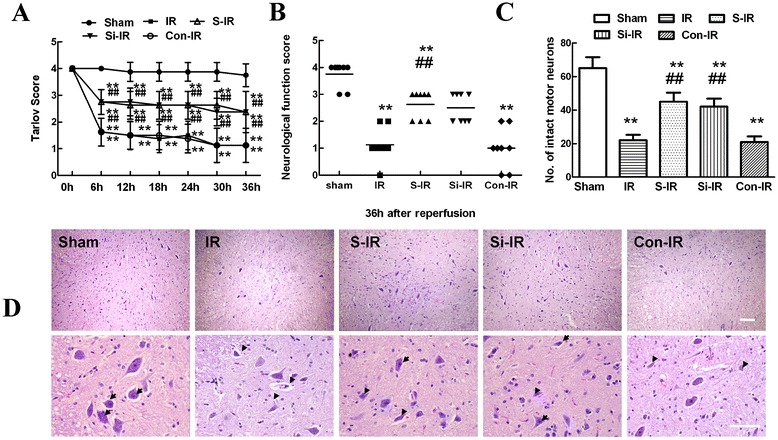
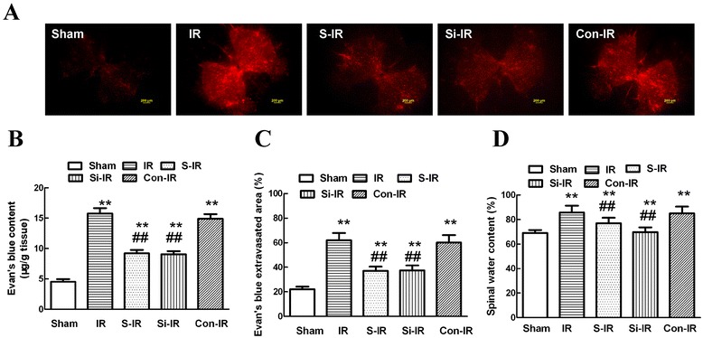
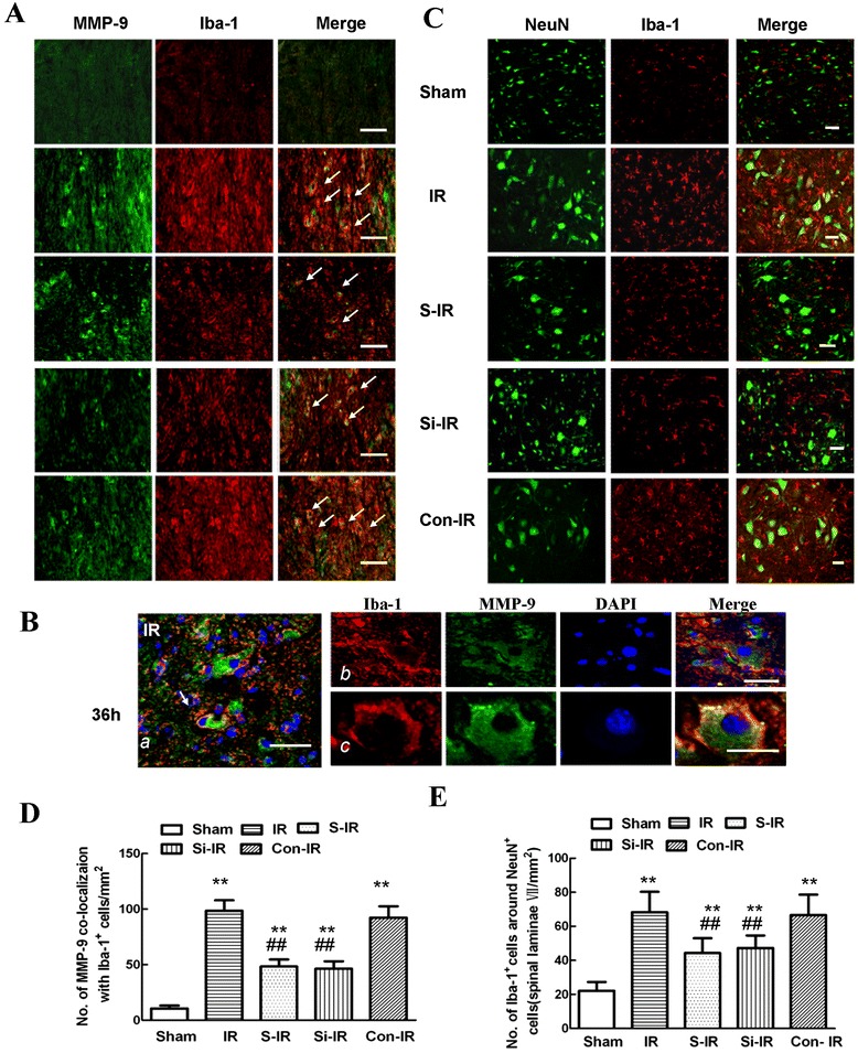
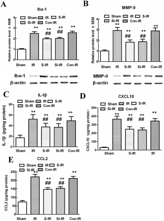
Results: The results showed significant upregulation of MMP-9 in rats with IR-induced inflammation of the BSCB compared to that of the sham group, manifested as dysfunctional BSCB with increased Evans blue extravasation and reduced expression of occludin protein. Increased MMP-9 expression was also observed to facilitate invasion and migration of activated microglia, imaging as high Iba-1 expression, clustered to neurons in the injured spinal cord, as shown by double immunofluorescence, and increased proinflammatory chemokine production (CXCL10, CCL2). Further, sevoflurane preconditioning markedly improved motor function by ameliorating neuronal apoptosis, as shown by reduced TUNEL-positive cell counts and expression of cleaved caspase-3. These protective effects were probably responsible for downregulation of MMP-9 and maintenance of normal expression of occludin protein indicating BSCB integrity from inflammatory damage, which was confirmed by decreased protein levels of Iba-1 and MMP-9, as well as reduced production of proinflammatory chemokines (CXCL10, CCL2) and proinflammatory cytokines (IL-1β). Intrathecal injection of specific siRNAs targeting MMP-9 had similar protective effects to those of sevoflurane preconditioning.

Conclusions: Preconditioning with 2.4% sevoflurane attenuated spinal cord IR injury by inhibiting recruitment of microglia and secretion of MMP-9; thus inhibiting downstream effects on inflammatory damage to BSCB integrity and neuronal apoptosis.


Figures

Figure 1
Figure 1
Effects of sevoflurane preconditioning on neurologic motor function and histologic assessment of the spinal cord after ischemia/reperfusion (IR) injury. (A) Neurological function scores were assessed at 6-h intervals during the 36 h observation using Tarlov scores after injury in three groups (n = 24). Neurological function scores ranged from 0 (paraplegia) to 4 (normal). Data are presented as the mean ± SEM. (B) Neurological function scores at 36 h after reperfusion in three groups. Each symbol represents data for one rat (n = 8, bar = median). (C) Number of intact motor neurons in the ventral gray matter (n = 8). (D) Representative sections of lumbar spinal cords in the ventral horn of gray matter stained with hematoxylin and eosin 36 h after reperfusion in three groups. Normal neurons exhibited a fine granular cytoplasm with Nissl substance (arrows), while dead neurons were identified by the presence of a diffuse cytoplasm without cellular structure and with extensive vacuolation of gray matter (arrowheads). Upper scale bar = 200 μm; lower scale bar = 100 μm. **P < 0.05 vs. sham group. ## P < 0.05 vs. IR group.
Figure 2
Figure 2
Effects of sevoflurane preconditioning on neuronal apoptosis after spinal cord ischemia reperfusion (IR) injury. (A) Representative micrographs of TUNEL staining (green) and DAPI (blue) in laminae IX of the ventral gray matter at 36 h after IR injury. Scale bars are 200 μm. (B) Representative immunohistochemical localization of neurons (NeuN; green) and cleaved-caspase-3 (red) in laminae IX of the spinal cord at 36 h after IR injury. Arrows delineate colocalization. Scale bars are 100 μm. Immunohistochemistry data showing that IR led to a decrease in neuronal number in the spinal ventral horn and an increased percentage of NeuN/cleaved-caspase-3-positive cells, suggesting the loss of neurons is partly a result of apoptosis. Pretreatment with sevoflurane and downregulation of MMP-9 by intrathecal injection of MMP-9 siRNA had neuroprotective effects reflected in decreased neuronal apoptosis. (C) Quantification of TUNEL-positive neurons in laminae IX of the ventral horn as averaged across three independent experiments. (D) Quantification of colocalized cells (cells with yellow signals) in laminae IX. (E) Representative western blot and quantitative protein analysis of cleaved caspase-3 in the spinal cord 36 h after surgery. Relative integral density values (IDVs) were calculated after normalizing to the sham group in each sample. All data are presented as mean ± SEM (n = 8 per group). **P < 0.05 vs. sham group; ## P < 0.05 vs. IR group.
Figure 3
Figure 3
Effects of sevoflurane preconditioning on blood-spinal cord barrier (BSCB) integrity after spinal cord ischemia reperfusion (IR) injury. (A) Effects of spinal cord IR injury on BSCB permeability measured by EB extravasation. Almost no red fluorescence was observed in the spinal cord of sham group rats 36 h after injury. Much more red fluorescence, especially in the gray matter, was observed in IR group rats 36 h after injury, whereas EB red extravasation was significantly less in the sevoflurane preconditioning group. Further, downregulation of MMP-9 by intrathecal injection with MMP-9 siRNA had similar neuroprotective effects on the integrity of the BSCB to those of sevoflurane preconditioning. (B) EB content of the spinal cord (μg/g). (C) Percentage of EB extravasated area. (D) Quantification of the water content of the spinal cord (edema). All data are presented as mean ± SEM (n = 8 per group). Scale bars are 200 μm. **P < 0.05 vs. sham group; ## P < 0.05 vs. IR group.
Figure 4
Figure 4
Effects of sevoflurane preconditioning on microglia activity, migration, and secretion of MMP-9 after spinal cord ischemia reperfusion (IR) injury. (A) Representative micrographs of the cellular location of MMP-9 (green) with antibodies against a microglial specific marker (Iba-1; red) 36 h after IR injury. Arrows delineate colocalization. A low magnification view showed fewer double-labeled cells in the spinal dorsal horn of rats preconditioned with sevoflurane or treated by intrathecal injection of MMP-9 siRNA. Arrows delineate colocalization. Scale bars are 100 μm. (B) Higher magnification image confirming that MMP-9 (with the identical fluorescence label) colocalized with Iba-1-positive cells 36 h after IR injury. Scale bars are 100 μm. (C) Distribution of Iba-1-positive cells in spinal lamina IX after IR injury. Representative micrographs showed that the IR-induced increase in Iba-1 immunoreactivity was prominently surrounded by NeuN-positive cells. Sevoflurane preconditioning prevented this redistribution. Similar neuroprotective effects were observed after downregulation of MMP-9 by intrathecal injection of MMP-9 siRNA. This suggests that the neuroprotective effects of sevoflurane preconditioning, in part, contributed to the inhibition of residual immune cell (microglia) activation and MMP-9 expression in the injured region. (D) Histogram for quantification of colocalized cells (cells with yellow signals). (E) Histogram for quantification of Iba-1-positive cells localized around NeuN-positive cells of lamina IX. All data are presented as mean ± SEM (n = 8 per group). **P < 0.05 vs. sham group; ## P < 0.05 vs. IR group.
Figure 5
Figure 5
Effects of sevoflurane preconditioning on proinflammatory chemokines and cytokines and neuronal apoptosis after spinal cord ischemia reperfusion (IR). (A-B) Representative western blot and quantitative protein analysis of Iba-1 and MMP-9 in the spinal cord 36 h after surgery. Relative integral density values (IDVs) were calculated after normalizing to the sham group in each sample. (C-E) Quantification of CXCL-10, CCL2, and IL-1β production in the spinal cord 36 h after IR injury, as assessed by ELISA. Sevoflurane preconditioning prevented IR-induced microglial activation and significant increases in the products of proinflammatory chemokines (CXCL10 and CCL2) and cytokines (IL-1β) 36 h after IR injury. Similar anti-inflammatory effects were observed after intrathecal injection of MMP-9 siRNA. All data are presented as mean ± SEM. **P < 0.05 vs. sham group; ##P < 0.05 vs. IR group.

References

    1. Kahn RA, Stone ME, Moskowita DM. Anesthetic consideration for descending thoracic aortic aneurysm repair. Semin Cardiothorac Vasc Anesth. 2007;11:205–223. doi: 10.1177/1089253207306098. - DOI - PubMed
    1. Aubé B, Lévesque SA, Paré A, Chamma E, Kébir H, Gorina R, Lécuyer MA, Alvarez JI, De Koninck Y, Engelhardt B, Prat A, Côté D, Lacroix S: Neutrophils Mediate Blood-Spinal Cord Barrier Disruption in Demyelinating Neuroinflammatory Diseases.J Immunol 2014, [Epub ahead of print]. - PubMed
    1. Li XQ, Lv H, Tan WF, Fang B, Wang H, Ma H: Role of the TLR4pathway in blood-spinal cord barrier dysfunction during the bimodal stage after ischemia/reperfusion injury in rats.J Neuroinflammation 2014, 11:62. - PMC - PubMed
    1. Li XQ, Wang J, Fang B, Tan WF, Ma H. Intrathecal antagonism of microglial TLR4 reduces inflammatory damage to blood-spinal cord barrier following ischemia/reperfusion injury in rats. Mol Brain. 2014;7:28. doi: 10.1186/1756-6606-7-28. - DOI - PMC - PubMed
    1. Wang XS, Fang HL, Chen Y, Liang SS, Zhu ZG, Zeng QY, Li J, Xu HQ, Shao B, He JC, Hou ST, Zheng RY. Idazoxan reduces blood–brain barrier damage during experimental autoimmune encephalomyelitis in mouse. Eur J Pharmacol. 2014;736:70–76. doi: 10.1016/j.ejphar.2014.04.034. - DOI - PubMed

Publication types

MeSH terms