Review
. 2014 Nov-Dec;3(6):465-87.
doi: 10.1002/wdev.152. Epub 2014 Sep 3.

Kidney: polycystic kidney disease

Affiliations
Review

Kidney: polycystic kidney disease

Binu M Paul et al. Wiley Interdiscip Rev Dev Biol. 2014 Nov-Dec.

Abstract

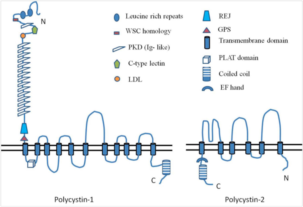
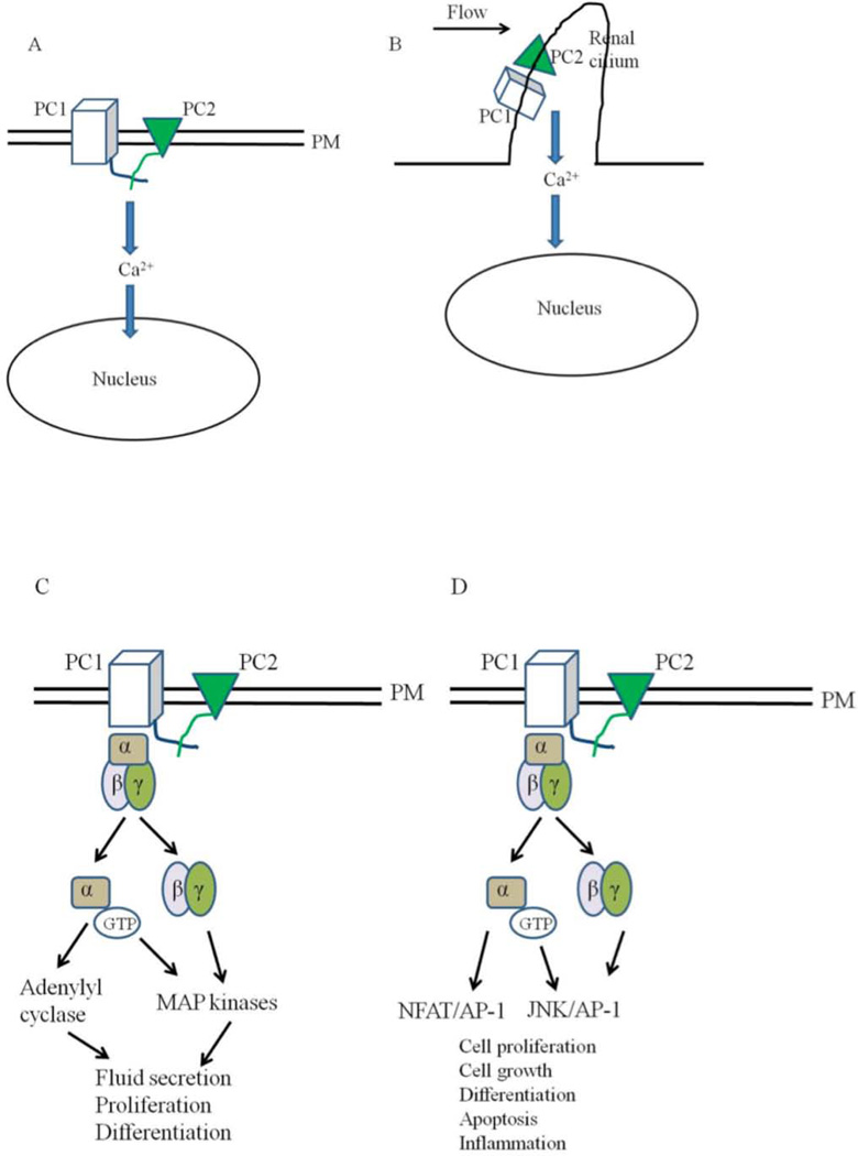
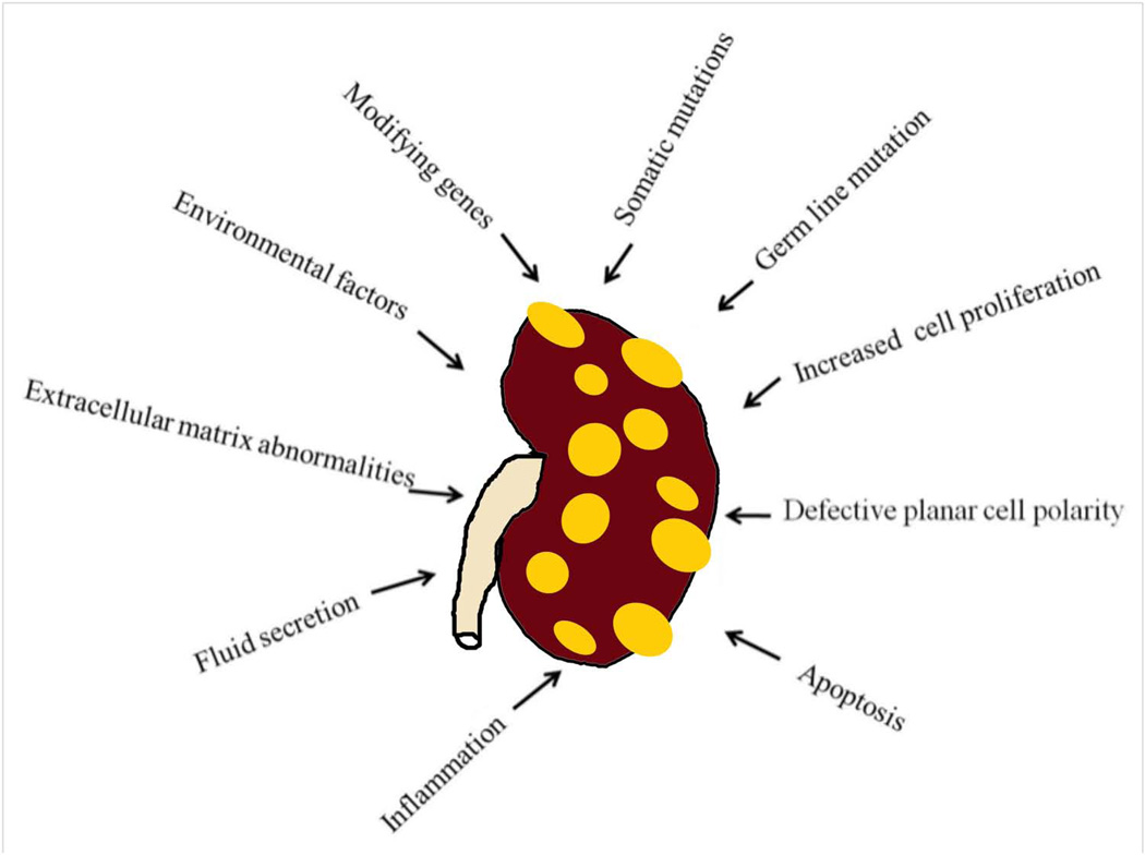
Polycystic kidney disease (PKD) is a life-threatening genetic disorder characterized by the presence of fluid-filled cysts primarily in the kidneys. PKD can be inherited as autosomal recessive (ARPKD) or autosomal dominant (ADPKD) traits. Mutations in either the PKD1 or PKD2 genes, which encode polycystin 1 and polycystin 2, are the underlying cause of ADPKD. Progressive cyst formation and renal enlargement lead to renal insufficiency in these patients, which need to be managed by lifelong dialysis or renal transplantation. While characteristic features of PKD are abnormalities in epithelial cell proliferation, fluid secretion, extracellular matrix and differentiation, the molecular mechanisms underlying these events are not understood. Here we review the progress that has been made in defining the function of the polycystins, and how disruption of these functions may be involved in cystogenesis.


Figures

Figure 1
Figure 1. Stages of kidney development in the mouse embryo
The pronephric duct arises from the intermediate mesoderm in the mouse embryo at E9.0. The nephric duct grows caudally until it reaches the cloaca. Mesonephric tubules can be seen at E10.0. The mesonephric tubules which are found more caudally, are more developed compared to the proximal ones. Posterior cells of the intermediate mesoderm specialize to form an aggregate called metanephric mesenchyme (red). The metanephric mesenchyme gives rise to all the segments of the nephron. The ureteric bud, an outgrowth from the nephric duct invades the MM by E10.5. The UB gives rise to the collecting system of the kidneys. The UB bifurcates and the induced mesenchyme, known as the cap mesenchyme (purple), surrounds the tips of the UB.
Figure 2
Figure 2. Stages of nephrogenesis
The adult metanephric kidney in mammals develops by a reciprocal interaction between the epithelial ureteric bud (blue) and the metanephric mesenchyme (purple). During the condensation stage, the loose mesenchymal cells of the MM condense around the tips of the epithelial UB. After the condensation stage, the induced mesenchyme undergoes a mesenchyme-to-epithelial conversion forms the comma and S-shaped structures. Later, the tubules elongate and the podocytes in the glomeruli fold to give rise to a mature nephron.
Figure 3
Figure 3. Structure of polycystins
Polycystin-1 has a large extracellular domain, 11 transmembrane domain and a short cytoplasmic tail. The coiled coil domain in the C-terminal end of PC-1 interacts with the C-terminal tail of Polycystin-2. Polycystin-2 has cytoplasmic N and C-terminus. Together, PC1 and PC2 mediate calcium entry into cells. (Modified from 2)
Figure 4
Figure 4. Models for the functions of polycystins
A: PC1 and PC2 are found on the plasma membrane (PM) in this model. Activation of PC1 leads to the activation of PC2 which leads to calcium influx and changes in gene transcription. B: Polycystins signaling through the primary cilia. Renal cilia bend in response to fluid flow. Bending of the cilia leads to the influx of calcium through polycystins. C and D: Polycystins regulating G-protein signaling. Binding of PC1 to heterotrimeric G proteins activates the Gα subunit and the release of the βγ subunits. This leads to the activation of adenylyl cyclases and MAP kinases which can affect several cellular processes such cell proliferation, fluid secretion and differentiation. G protein signaling through polycystins can also affect the JNK/AP-1 and NFAT/AP-1 pathways. E: Polycystins signaling through the JAK-STAT signaling pathway. E: Membrane anchored PC1binds to JAK2 and activates it which then phosphorylates STAT3. F: PC1 C tail can translocate to the nucleus and coactivate STAT1, 3 and 6 which were already activated independently through cytokine signaling. Once in the nucleus, STAT transcription factors affect the expression of genes required for cell proliferation, cell growth, differentiation and apoptosis. G: Polycystins regulating the JAK-STAT signaling pathway. PC1, in a reaction requiring PC2, activates JAK2 which leads to the phosphorylation of STAT1. Phosphorylated STAT1 translocates to the nucleus and activates the cyclin-dependent kinase inhibitor, p21. Activation of p21 results in the subsequent inhibition of cdk2 and cell cycle arrest.
Figure 5
Figure 5. Potential mechanisms of cystogenesis and disease progression in ADPKD

References

    1. Grantham JJ. Polycystic kidney disease: huge kidneys, huge problems, huge progress. Trans Am Clin Climatol Assoc. 1997;108:165–170. discussion 70-2. - PMC - PubMed
    1. Igarashi P, Somlo S. Genetics and pathogenesis of polycystic kidney disease. J Am Soc Nephrol. 2002 Sep;13(9):2384–2398. - PubMed
    1. Harris PC, Torres VE. Genetic mechanisms and signaling pathways in autosomal dominant polycystic kidney disease. J Clin Invest. 2014;124:2315–2324. - PMC - PubMed
    1. Gallagher AR, Germino GG, Somlo S. Molecular advances in autosomal dominant polycystic kidney disease. Adv Chronic Kidney Dis. 2010;17:118–130. - PMC - PubMed
    1. Hou X, Mrug M, Yoder BK, Lefkowitz EJ, Kremmidiotis G, D'Eustachio P, et al. Cystin, a novel cilia-associated protein, is disrupted in the cpk mouse model of polycystic kidney disease. J Clin Invest. 2002 Feb;109(4):533–540. - PMC - PubMed

Publication types

MeSH terms

LinkOut - more resources